Skin tumours: find your pathway
Basal cell carcinoma, cutaneous squamous cell carcinoma, Merkel cell carcinoma and rare skin tumours have different biology and different treatment pathways. Start with your situation, not with complex terminology.
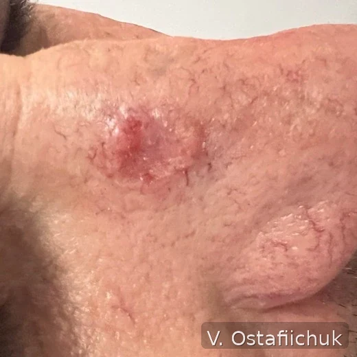
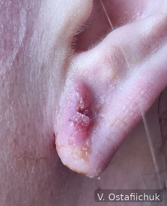
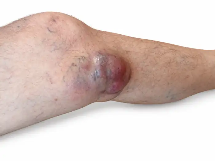
Stage groups, TNM categories, translated definitions and site-specific limits.
What brought you here?
You do not need to know the stage, risk group or treatment terminology. Choose the situation that most closely matches yours.
When biopsy is needed and why a photograph or dermoscopy does not replace histology.
02My pathology report says BCCWhat basal cell carcinoma means, when standard excision may be enough and when more complex margin control is considered.
03My pathology report says cSCCWhy risk factors, depth, nerves, anatomic site and regional nodes matter in cutaneous squamous cell carcinoma.
04I was told I have Merkel cell carcinomaA separate pathway: pathology confirmation, staging, nodes, surgery, radiotherapy and systemic treatment.
05I was advised to have Mohs or margin-controlled surgeryWhy margins may be checked in stages and why this matters particularly at anatomically complex sites.
06I will need reconstruction after tumour removalDirect closure, local flaps, grafts and planning around functionally important anatomy.
07I have an enlarged lymph nodeWhen ultrasound, CT or needle biopsy may be needed and why the logic differs between cSCC and Merkel.
08I have completed treatmentHow the scar, skin, nodes and new lesions are monitored and when additional imaging may be appropriate.
If you remember only three things
This is enough to avoid getting lost at the start.
BCC, cSCC, Merkel and rare skin tumours differ in risk, staging and treatment. The pathology report is the first key document.
Eyelids, nose, lip, ear, scalp, hand and foot may require different thinking about margins, nodes and reconstruction.
The tumour must first be adequately controlled, but function and defect closure should be considered before the operation begins.
Which skin tumour are we talking about?
The tumour type defines the basic pathway. Site, pathological risk features, nodal status and disease extent then refine the plan.
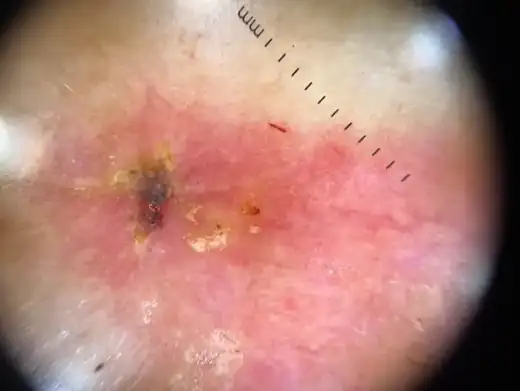
From local lower-risk BCC to recurrent tumours and anatomically complex facial sites.
Depth, differentiation, PNI, anatomic site and nodes can substantially alter management.
Biopsy and IHC, distinct nodal staging, surgery, radiotherapy and immunotherapy pathways.
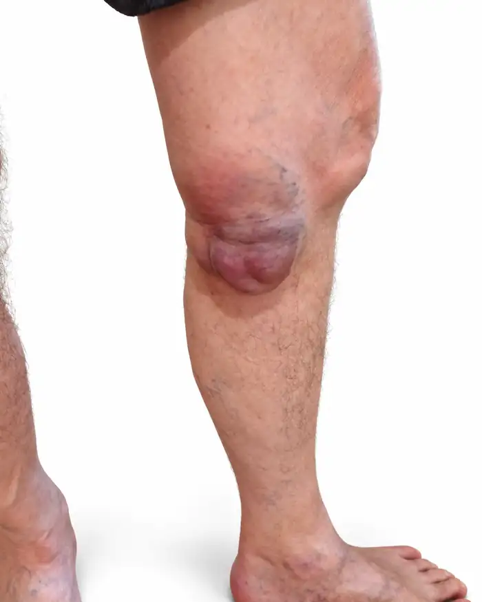
Sebaceous carcinoma, EMPD, angiosarcoma, porocarcinoma, adnexal carcinomas and other rare diagnoses.
From suspicion to follow-up
Not every patient needs every step. But the sequence helps ensure that important decisions are not missed.
Clinical review, dermoscopy and assessment of anatomic site.
02BiopsyObtain adequate tissue for diagnosis.
03PathologyTumour type, risk features, margins and other key parameters.
04Risk and stagingNodal assessment and imaging are needed in selected patients.
05Tumour controlSurgery, Mohs, radiotherapy or another method where appropriate.
06ReconstructionClose the defect without compromising oncologic treatment.
07Follow-upScar, skin, nodes, self-examination and prevention.
From diagnosis to reconstruction
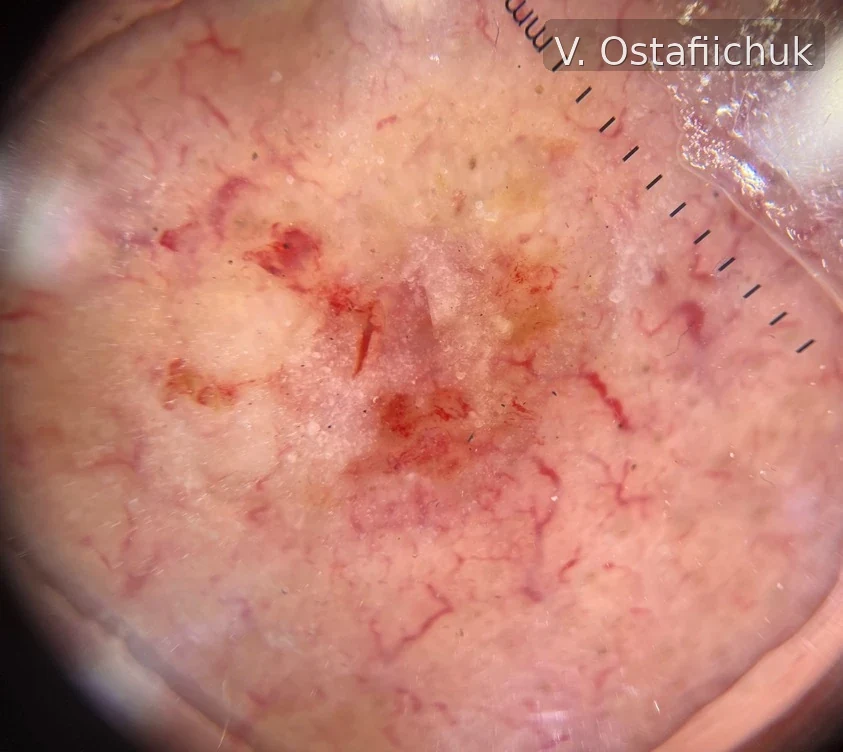
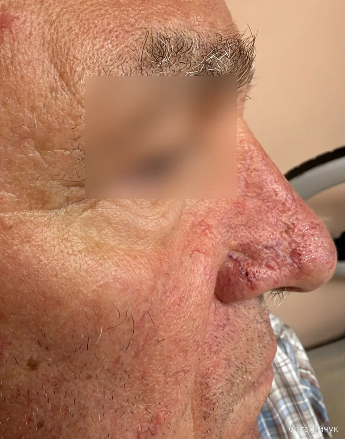
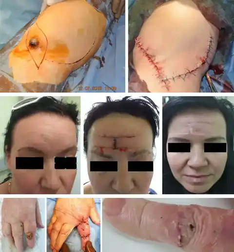
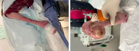
Images support the pathway and explain clinical reasoning, but they are not intended for remote self-diagnosis.
Patient guides
Short pages for a specific situation - before biopsy, after surgery or during follow-up.
What tissue the pathologist needs and why biopsy technique depends on the clinical situation.
Open the guide →Tumour type, margins, PNI/LVI and other parameters that may change the next decision.
Patient explanation →What may be expected early and which findings should prompt contact with the treating team.
Postoperative guide →When ultrasound, needle biopsy or additional imaging may be appropriate.
Nodal assessment pathway →Monitoring the scar, entire skin, regional nodes and new lesions.
Follow-up plan →UV exposure, clothing, shade, SPF and practical protection for scars and skin.
Practical sun protection →Eyelid, nose, ear, lip and scalp
The same histology may require a different strategy depending on site, function and reconstructive options.
Margin control, eyelid function and differential diagnosis, including sebaceous carcinoma.
BCC at complex sites →Mohs and local flaps can combine tumour control with preservation of contour.
Author clinical case →For cSCC and selected rare tumours, anatomic site changes staging and operative planning.
cSCC pathway →Rare tumours should not disappear under the label “skin cancer”
Merkel, sebaceous carcinoma, EMPD, angiosarcoma, porocarcinoma and other adnexal tumours require their own pathological and clinical logic.
For clinicians
BCC/cSCC risk stratification, pathology, margins, PNI/LVI, imaging, nodal staging, SLNB in Merkel, adjuvant and systemic decisions, rare skin tumours and the contemporary 2024-2026 evidence base.
The patient level deliberately simplifies terminology. Full risk-stratification systems, staging, pathology parameters and professional algorithms are separated into the clinician section.