Bone tumours.
Understand. Preserve. Restore.
From a concerning X-ray finding to expert histopathology, multidisciplinary planning and reconstruction. A patient pathway, real clinical imaging and an evidence-based clinician layer.
What brought you here?
You do not need to know the pathology yet. Choose the decision you need to make next.
The diagnostic sequence
Radiographs are often first; CT and MRI are added according to the lesion.
Local extent, the chest and other areas are assessed according to the suspected diagnosis.
The biopsy track is placed within the future resection rather than selected in isolation.
Oncological removal and restoration of bone or joint function are planned together.
When not to delay assessment
Persistent or night bone pain, swelling, pain after minor loading, possible pathological fracture or progressive loss of function should be investigated. A normal X-ray does not always exclude serious disease. Seek urgent care if you cannot bear weight or a fracture is suspected.
Explore the centre
Real documented cases
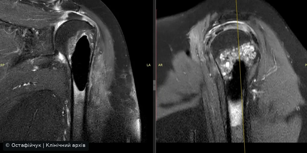
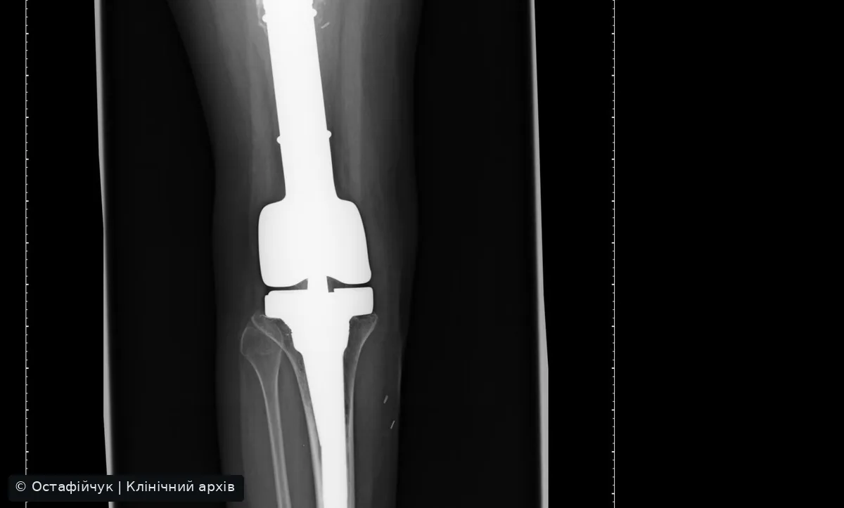
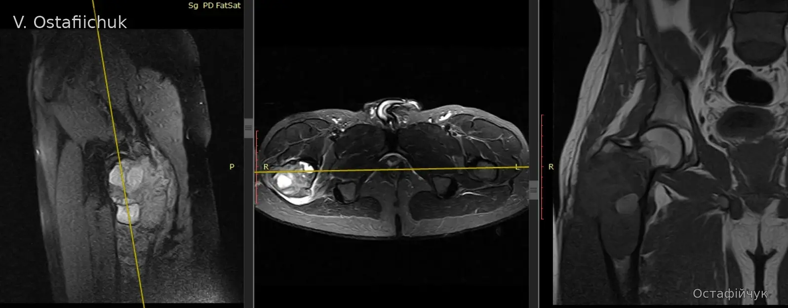
Proximal humerus chondrosarcomaMRI and documented resection, cryotherapy and bone cement →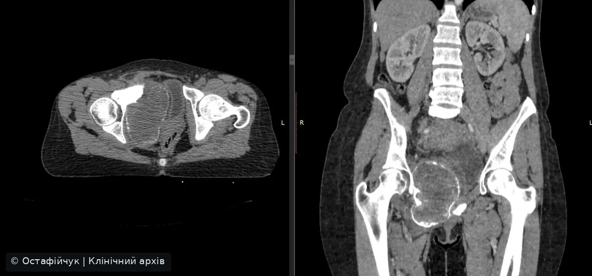 Giant cell tumour of the pubic boneBiopsy, denosumab, CT review and resection planning →
Clinical media are de-identified. Where pathology is not supplied, the imaging atlas describes anatomy without asserting a diagnosis.
Common questions
Does every bone lesion require surgery?
No. Some benign findings need surveillance only; suspicious or aggressive lesions may require specialist imaging and planned biopsy.
Can a biopsy be performed before specialist review?
Suspected bone sarcoma should first undergo appropriate MRI and specialist biopsy tract planning.
Is limb-sparing surgery an option?
Often, yes. Feasibility depends on pathology, extent, achievable margins and safe functional reconstruction.
References: UK Bone Sarcoma Guidelines 2025, ESMO Bone Sarcoma Guidelines.
TNM, grade, pelvic/spinal exceptions and why not every bone lesion receives a malignant bone sarcoma stage.
Primary bone tumour staging
Four components describe the anatomical extent and histological grade, but a stage group cannot be inferred from size alone. Confirm the diagnosis and the applicable anatomical system first.
Important: pelvic and spinal primary bone tumours have specific T categories without the usual prognostic stage groups IA-IVB. Giant cell tumour and benign lesions do not receive a malignant bone sarcoma AJCC stage.
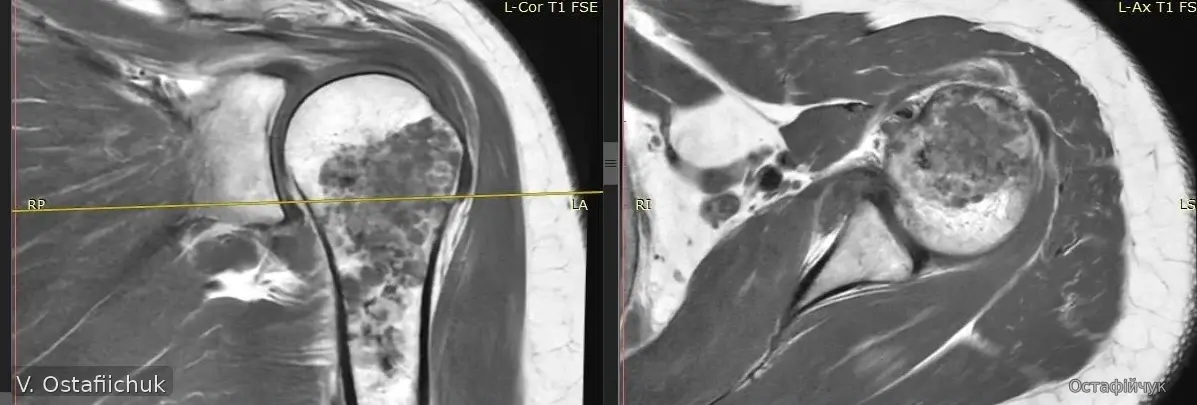
Open the full AJCC 8 clinician staging guide →MRI examples
Short operative clip

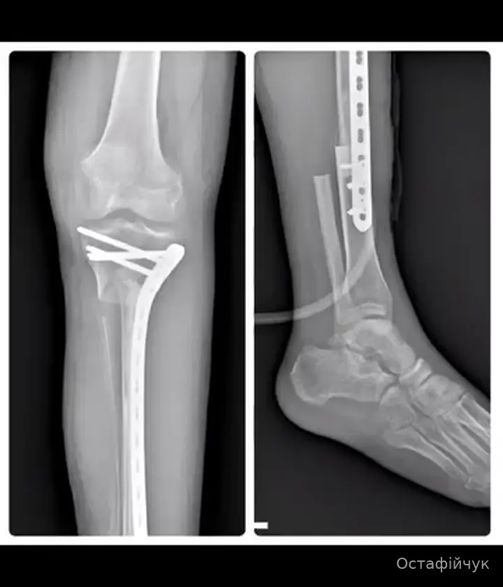
Clinical material from the author's archive, published with confirmed consent.