Soft-tissue sarcoma: find your pathway
If a soft-tissue mass has been found, the first step is not always removal. The safer pathway is imaging first, then a planned biopsy, an exact pathological diagnosis and only then definitive treatment.
Stage groups, TNM categories, translated definitions and site-specific limits.
Where are you in the pathway?
You do not need to know the stage or medical terminology. Choose the situation closest to yours.
When it needs specialist assessment and what should happen before biopsy.
02A biopsy is being plannedWhy MRI should usually come first and why biopsy trajectory matters.
03The tumour was already removedWhat happens next: MRI of the bed, pathology review and MDT planning.
04Sarcoma is confirmedWhat type it is, how aggressive it appears and whether resection margins are clear.
05Surgery or radiotherapy is being plannedWhen radiotherapy is given before surgery, when after, and how this affects wound healing.
06Do I need drug treatment?Why drug choice depends on the exact sarcoma subtype and clinical setting.
07Lung nodules were foundWhen surgery, stereotactic radiotherapy, ablation or systemic treatment may be discussed.
08I completed treatmentHow the operative site, lungs, function and late treatment effects are monitored.
If you remember only three things
These principles help prevent the most common early mistakes.
A suspicious deep or large lesion should usually be imaged before tissue is taken.
The biopsy trajectory is planned so it can be removed with the tumour during definitive surgery.
Pathology, immunohistochemistry and selected molecular tests determine whether surgery, radiotherapy or systemic treatment is appropriate.
Diagnosis is built from several layers
No single MRI and no single slide provides the whole answer. Anatomy, tissue morphology and tumour biology need to be interpreted together.
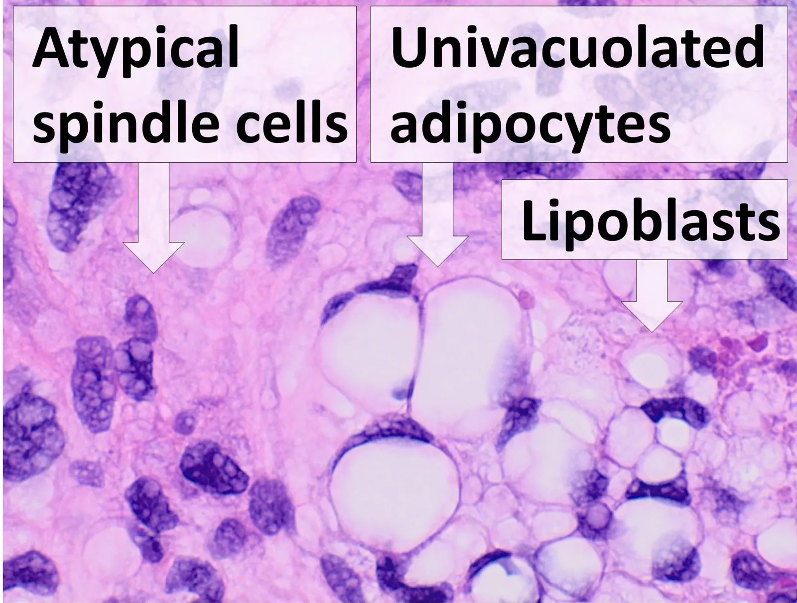
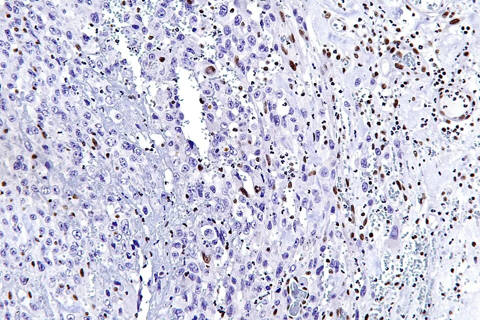
Open-license micrographs are mirrored locally during the site build. Source and license are retained for every asset; copyrighted journal figures are not copied without reuse permission.
Why a specialized sarcoma treatment unit matters
Sarcoma outcomes depend on more than the operation itself. Imaging before biopsy, biopsy trajectory, expert pathology, MDT review, resection planning, reconstruction, radiotherapy and systemic treatment work best as one coordinated pathway.
These are large prospective/registry observational data rather than randomized comparisons. The appropriate interpretation is that care in specialized sarcoma centers is associated with better outcomes. Sources: GEIS 2018, NETSARC 2019.
From suspicion to surveillance
Each step answers a different question. The order matters.
What actually determines risk?
Sarcoma stage is not based on size alone. Anatomical site and precise histology also matter.
Larger and deeper tumours generally require more complex local planning.
For many adult STS, FNCLCC combines differentiation, mitotic activity and necrosis.
Nodal spread is uncommon for most STS, while the lungs are a frequent site of distant metastasis.
Staging rules differ for extremity/trunk, retroperitoneal and other sarcoma sites.
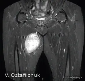
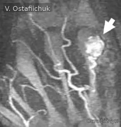
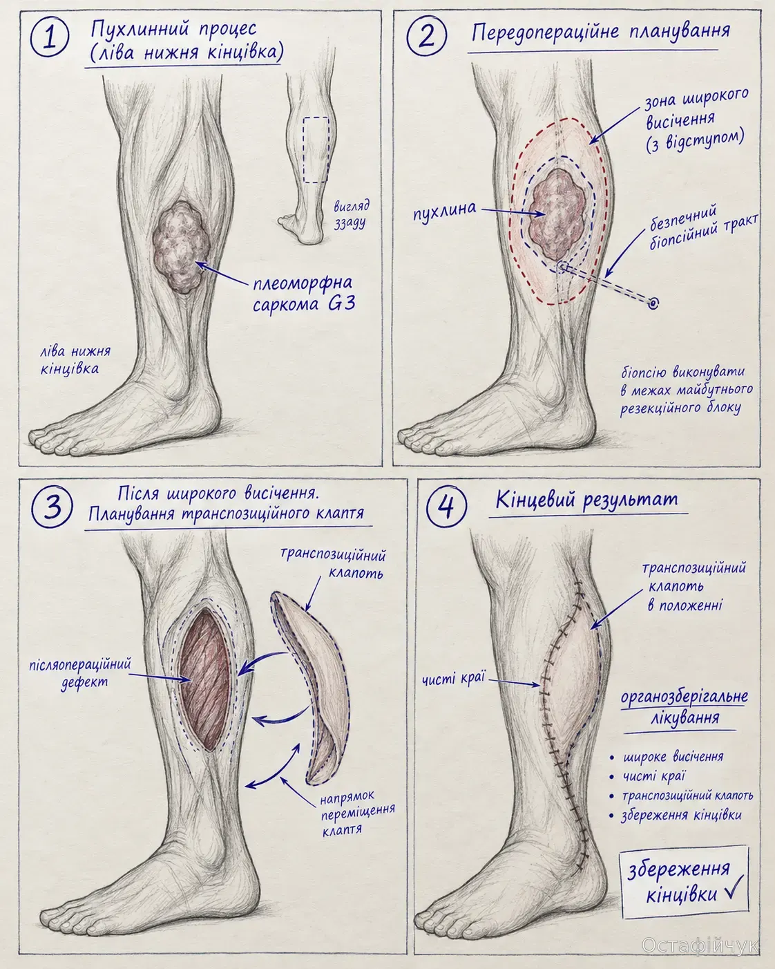
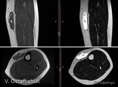
Imaging, vessels, surgery and reconstruction
These materials explain treatment planning; they are not intended for remote self-diagnosis.
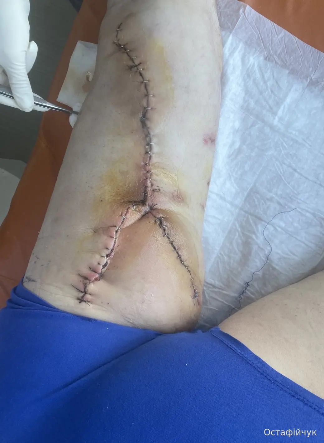
Short preoperative-planning video
Clinical material is de-identified and comes from the author’s clinical archive approved for educational publication.
Ministry of Health Standard No.1434 remains the national framework for extremity and trunk STS and requires specialist multidisciplinary care, morphological confirmation, MRI and staging imaging. DFSP has a separate national protocol, No.828, since 2026.
For clinicians
AJCC 8, biopsy planning, pathology and molecular confirmation, surgical margins, radiotherapy, histology-adapted systemic treatment, pulmonary metastases and current 2025-2026 guidance.
The patient layer deliberately uses simpler terminology. Detailed AJCC staging tables, professional abbreviations and evidence summaries are kept in the clinician section.