Follow-up after sarcoma treatment: what to monitor and for how long
A single clear scan does not end the sarcoma pathway. Follow-up should look for local recurrence, metastatic disease and late treatment effects while supporting long-term function.
What follow-up is trying to detect
Local recurrence
Clinical review, patient self-awareness and local MRI or ultrasound when appropriate. MRI is particularly valuable for deep tumours and complex postoperative beds.
Metastatic disease
The lungs are a common metastatic site for many STS subtypes, so regular chest assessment remains an important part of surveillance.
Late treatment effects
Limb function, oedema, radiation fibrosis, neuropathic pain, contracture, anthracycline cardiotoxicity and fertility issues may all require long-term attention.
A practical timeline: Ukraine and international guidance
Ukrainian Standard No.1434 suggests approximately every 3 months. SEOM-GEIS recommends every 3-4 months for intermediate/high-risk disease.
This is a common risk-adapted pattern once the highest early-risk period has passed.
International guidance often moves toward annual review. Ukrainian guidance explicitly recognises late recurrence.
The Ukrainian standard advises discussing ongoing follow-up even beyond 10 years.
What may be included
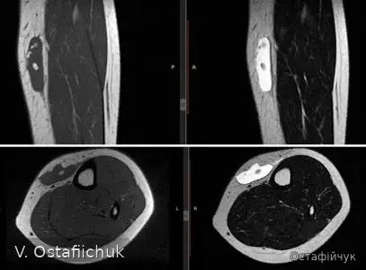
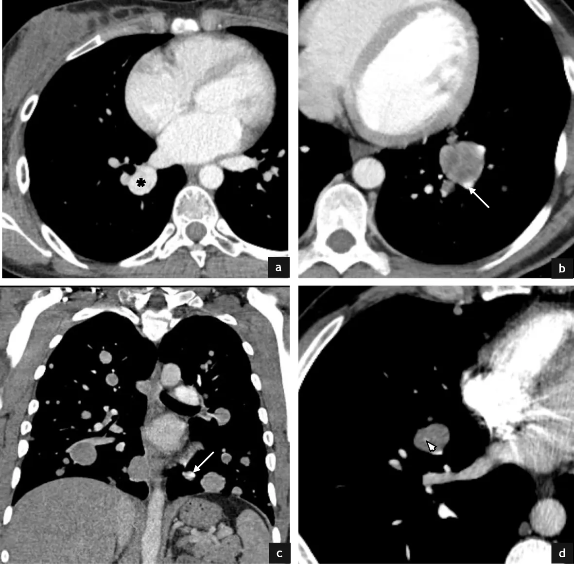
Clinical review and MRI adapted to risk, anatomy and the postoperative bed.
Chest CT or another risk-adapted chest-imaging strategy over time.
Oedema, fibrosis, mobility, pain, neurological symptoms and rehabilitation needs.
Ukrainian guidance includes chest CT and allows alternation with chest radiography after year 3 in selected situations. International practice also adapts chest imaging to risk.
Especially useful for deep tumours or a complex operative bed. ESSR supports risk- and subtype-adapted imaging rather than one universal schedule.
The scar, operative region, function, swelling, pain and neurological symptoms should be considered together with imaging.
Some sarcomas have unusual metastatic patterns, so the surveillance plan may extend beyond “chest CT plus local MRI”.
When not to wait for the next scheduled visit
- a new or enlarging lump near the scar;
- new persistent pain or swelling;
- loss of limb function;
- shortness of breath, haemoptysis or other new respiratory symptoms;
- new neurological symptoms or other changes not explained by expected postoperative recovery.
Standard No.1434 uses a lifelong follow-up principle and specifically addresses late recurrence and late treatment effects. This is important because “five years have passed” does not automatically mean surveillance should stop for every sarcoma.
Clinical images on this page are de-identified materials from the author’s clinical archive that were already approved for educational publication. No identifying DICOM overlays or patient identifiers are displayed.
The pathway matters
Follow-up should also be individualized: the risks of local recurrence, pulmonary metastasis and late toxicity differ between sarcoma entities. Continuity in a specialized sarcoma unit reduces fragmented surveillance.
GEIS 2018: 3-year overall survival 82% in reference centers vs 70.4% in local hospitals. NETSARC 2019: surgery in a reference center was associated with better overall survival (HR 0.681). Observational data. GEIS · NETSARC.