What should a sarcoma pathology report contain?
The single word “sarcoma” is not enough to plan treatment. Subtype, grade, resection margins and molecular features can change the next step.
A diagnosis according to the current WHO classification, rather than only a broad label such as “spindle-cell sarcoma” when a more specific entity can be established.
Grade integrates differentiation, mitotic activity and necrosis. Not every sarcoma subtype is appropriately graded with FNCLCC.
These parameters contribute to biological risk assessment and, for applicable entities, to histological grading.
Needed when morphology alone is insufficient, when a defining molecular event is expected, or when the result may open a treatment option.
For resection specimens, the maximum tumour size, tissue planes and involvement of fascia, muscle, vessels, nerves or other structures matter.
Not only R0/R1/R2, but which margin is closest, the microscopic distance and whether an anatomic barrier lies between tumour and margin.
What sarcoma can look like under the microscope
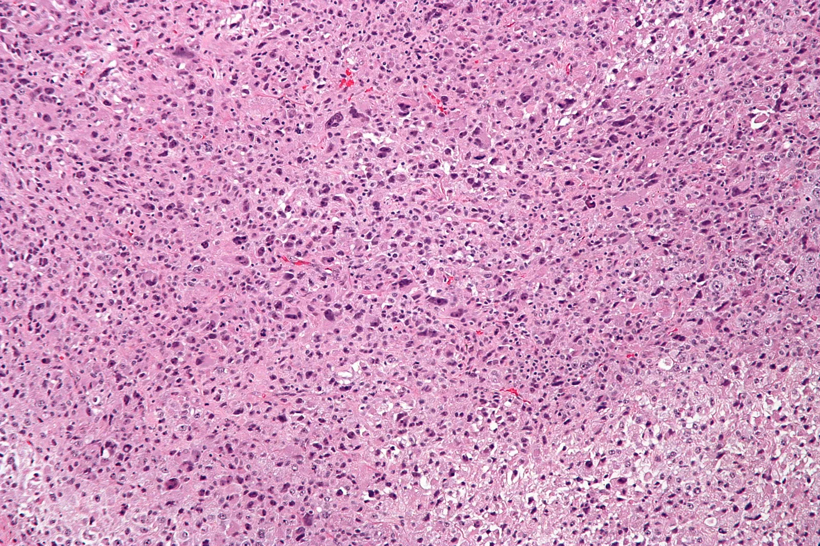
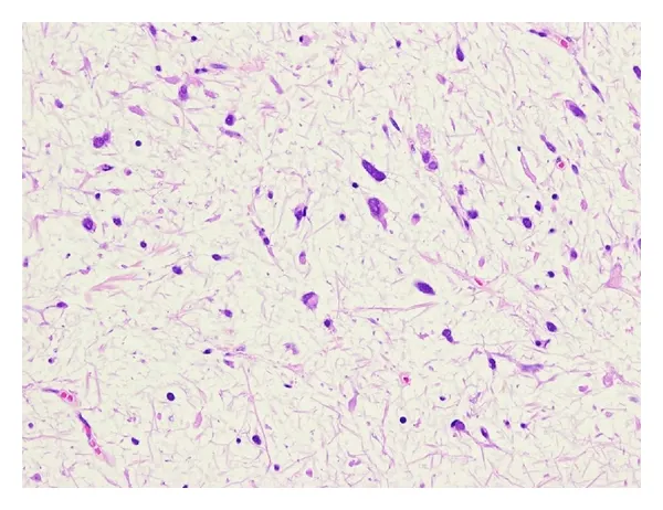
Micrographs are not a self-diagnostic tool. They illustrate why sarcoma pathology is specialised: different entities can look very different, while others overlap and require additional tests.
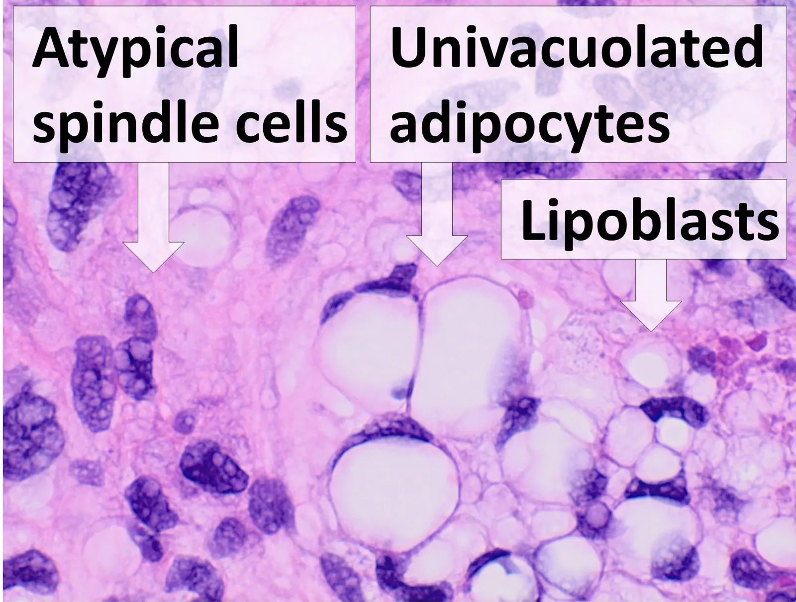
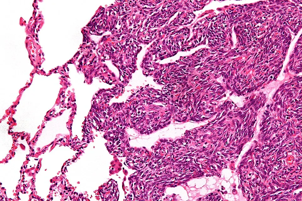
Open-license images are shown with author and licence information and do not represent a patient from this website.
R0, R1 and R2 in practical terms
Tumour does not reach the assessed resection margin.
Tumour cells reach the margin, although no gross residual tumour is visible.
Visible tumour remains after surgery.
Microscopically negative margins
No tumour cells are seen at the assessed margins. This does not mean zero recurrence risk; subtype, grade, size and other factors still matter.
Microscopically positive margin
Tumour reaches a resection margin. Management may involve re-excision, radiotherapy or another MDT strategy depending on anatomy and tumour biology.
Macroscopic residual tumour
Visible tumour remains after surgery. This is a different clinical situation requiring reassessment of resectability and multimodality treatment.
The word “sarcoma” can hide very different morphology
Why grade must be interpreted together with subtype
FNCLCC is useful for many adult-type soft-tissue sarcomas, but there are important exceptions. DFSP, atypical lipomatous tumour/well-differentiated liposarcoma, epithelioid sarcoma, alveolar soft-part sarcoma and other entities follow different grading or risk logic. The final risk should therefore never be inferred from one grade number alone.

When expert review is especially valuable
- the diagnosis is broad or uncertain;
- clinical and MRI findings do not fit the pathology;
- the subtype is rare;
- the result will determine a major operation or amputation;
- molecular testing may be required;
- the tumour was already removed outside a sarcoma center.
National Standard No.1434 requires morphological confirmation before special treatment and adequate tissue for histology and immunohistochemistry. Complex cases are best reviewed in a center where pathology is integrated with imaging, surgery and MDT planning.
Clinical images on this page are de-identified materials from the author’s clinical archive that were already approved for educational publication. No identifying DICOM overlays or patient identifiers are displayed.
Questions to ask after receiving the report
Has the exact subtype been established?
If not, ask whether there is enough tissue and whether additional IHC or molecular testing could clarify the diagnosis.
Is FNCLCC applicable to this subtype?
Not every sarcoma should be graded with FNCLCC. The grading system and its relevance should be clear.
What does my resection margin mean?
R1 does not automatically mean amputation, and R0 does not mean zero recurrence risk. Anatomy and tumour biology still matter.
Is molecular confirmation needed?
It is particularly useful when the diagnosis is uncertain, when a defining genetic event is expected or when the result could alter treatment.
The pathway matters
In rare morphology, an error in subtype or grade can alter the entire treatment plan. In a specialized sarcoma treatment unit, pathology is interpreted together with imaging, clinical context and the question that molecular testing needs to answer.
GEIS 2018: 3-year overall survival 82% in reference centers vs 70.4% in local hospitals. NETSARC 2019: surgery in a reference center was associated with better overall survival (HR 0.681). Observational data. GEIS · NETSARC.