When soft-tissue sarcoma is suspected
A safe sequence of imaging, biopsy and specialist planning.
Do not rush to removal
Most soft-tissue masses are benign, but an unplanned excision of a sarcoma can complicate definitive surgery.
Imaging first
The imaging modality depends on anatomical site and clinical context. MRI is commonly used for local assessment of extremity and trunk lesions.
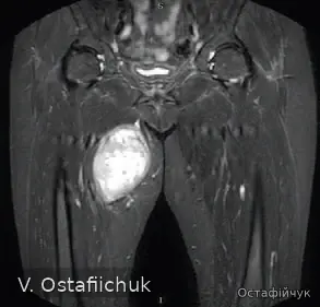
Clinical images from the author's archive, published with patient consent.
Planned biopsy
When tissue diagnosis is required, the approach and biopsy tract should be planned with the surgical team because the tract may need to be removed during definitive surgery.
Multidisciplinary review
Pathology, local imaging, staging and treatment options should be brought together before a final plan is made.
Reviewed September 2026. General information only; individual decisions require clinical assessment.
The pathway starts before surgery
The benefit of specialist care starts before the first intervention: correct imaging, a planned biopsy and MDT review before surgery reduce the risk of unnecessary or repeat procedures.
GEIS 2018 reported 3-year overall survival of 82% in reference centers vs 70.4% in local hospitals. NETSARC 2019 found surgery in a reference center independently associated with better overall survival (HR 0.681). These are observational data, not randomized comparisons. GEIS · NETSARC.