Clinical case: proximal humerus chondrosarcoma
A documented historical case, with unknowns clearly identified.
Preoperative imaging
MRI demonstrates a proximal humeral intramedullary lesion. Imaging needs correlation with radiographs, CT and pathology because tumour grade cannot be inferred from appearance alone.
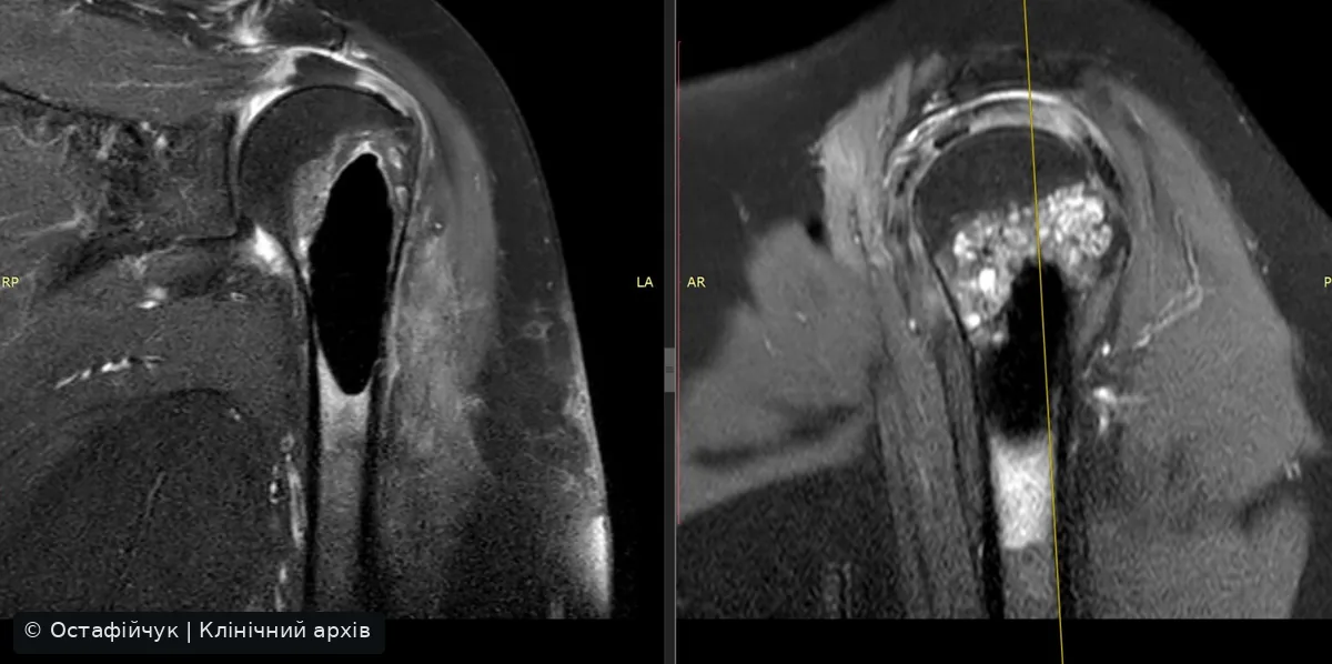
Documented operation
The archival presentation describes marginal proximal humeral resection, curettage, cryotherapy and bone cement reconstruction. This is an account of one operation, not a universal recommendation for chondrosarcoma.
Why this case matters
It illustrates the need to distinguish cartilaginous lesion biology, choose appropriate margins, consider local adjuvants and preserve shoulder function without compromising cancer care.
What is not available
No TNM stage, grade, R0/R1 status or recurrence-free outcome is inferred without corresponding source documentation.
Clinical sources
- UK guidelines for the management of bone sarcomas, 2025
- ESMO-EURACAN-GENTURIS-ERN PaedCan, Bone Sarcoma CPG
- NCI Osteosarcoma Treatment (PDQ)
- NCI Ewing Sarcoma Treatment (PDQ)
Clinical images and videos from the author’s archive. Patient identifiers have been removed. Diagnoses are stated only where supported by source records. This information does not replace an individual multidisciplinary decision.