Clinical imaging atlas
Bone tumour imaging atlas: MRI, CT and video
Real clinical images show how the team plans investigations and surgery.
How to use this atlas: histology is stated only when confirmed by a source record. Other items demonstrate imaging patterns, not diagnoses. Different images may belong to different patients.
Bone lesions, CT, MRI and reconstruction
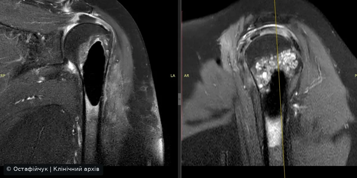
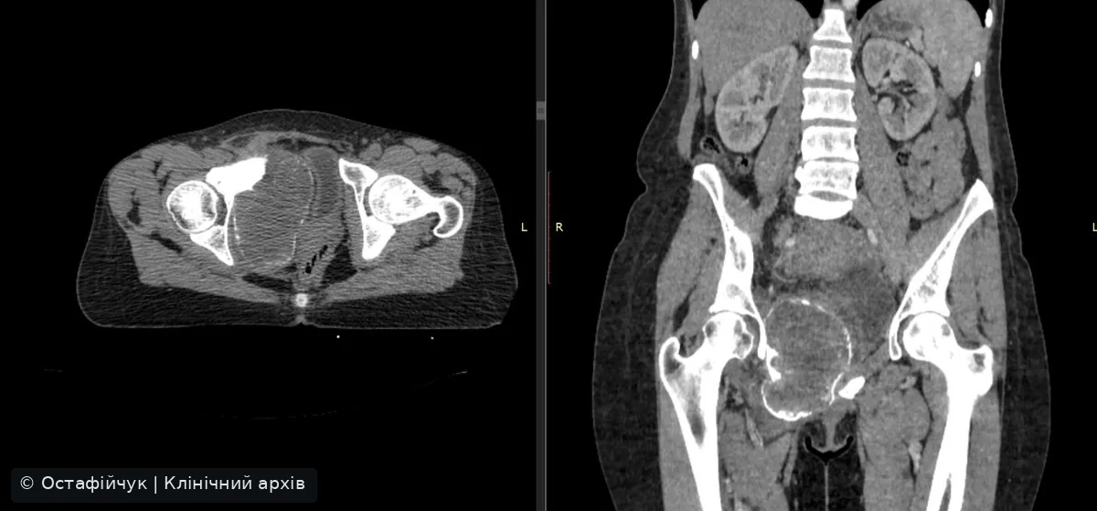
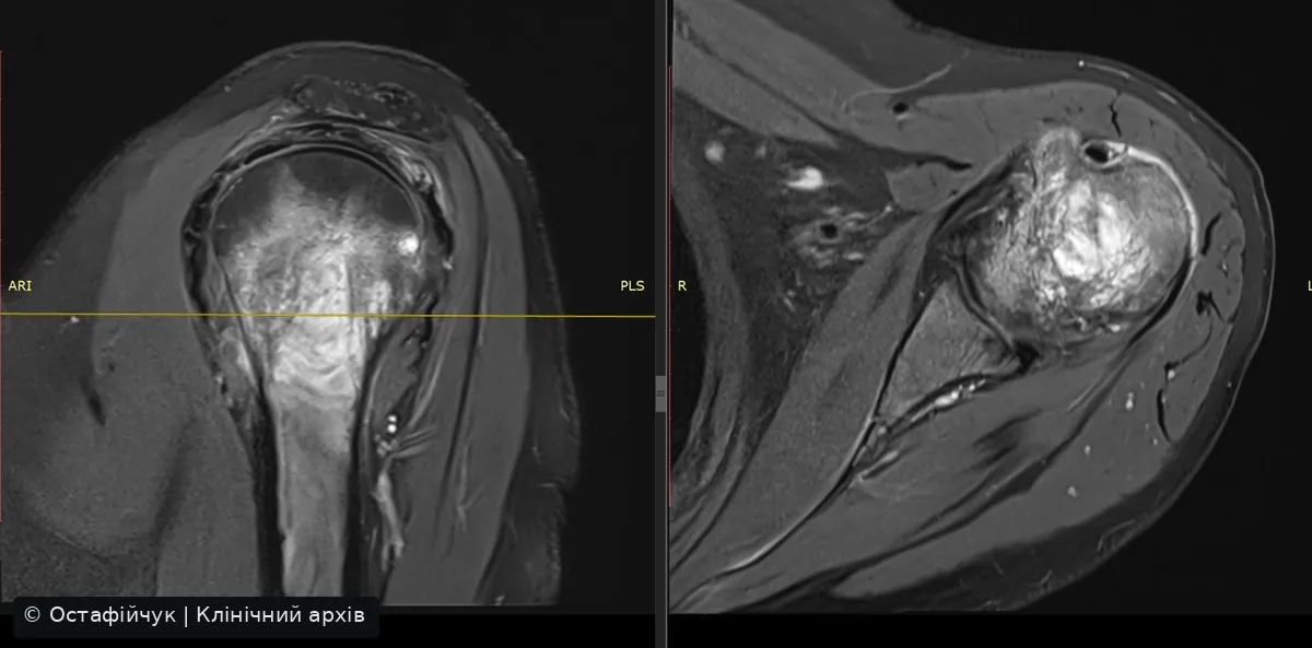
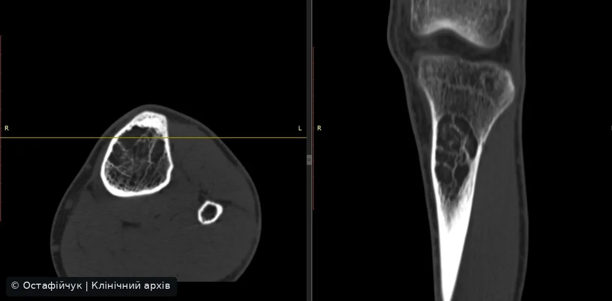
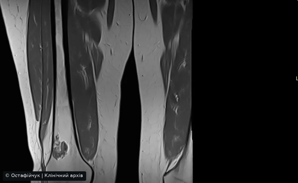
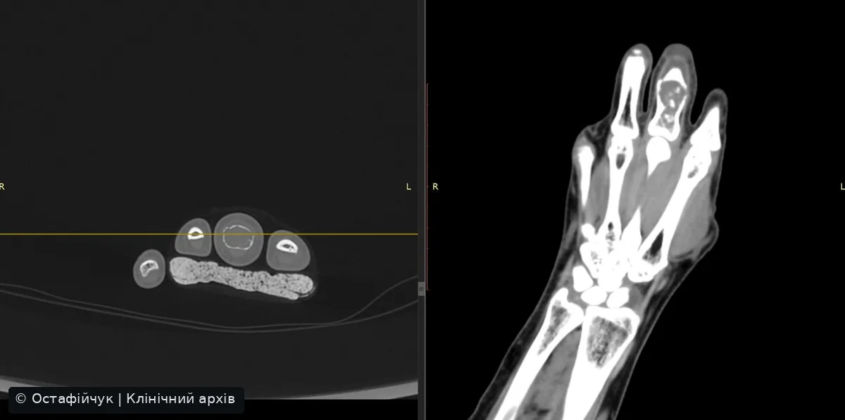
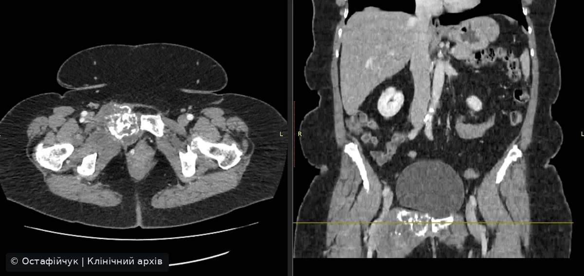
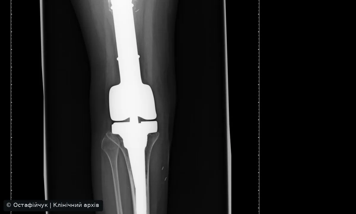
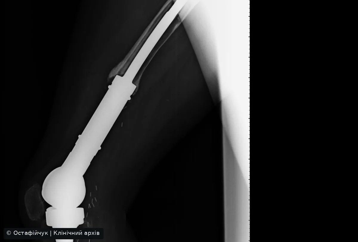
MRI clips: the value of scrolling through slices
Short clips from separate studies in the clinical archive. They illustrate changing anatomy through sequential slices and are not sufficient for diagnosis. Histology has not been linked to these clips.
Differential diagnosis: soft-tissue lesions
A key distinction is whether a mass arises from bone or adjacent soft tissues. This affects biopsy planning and surgery.
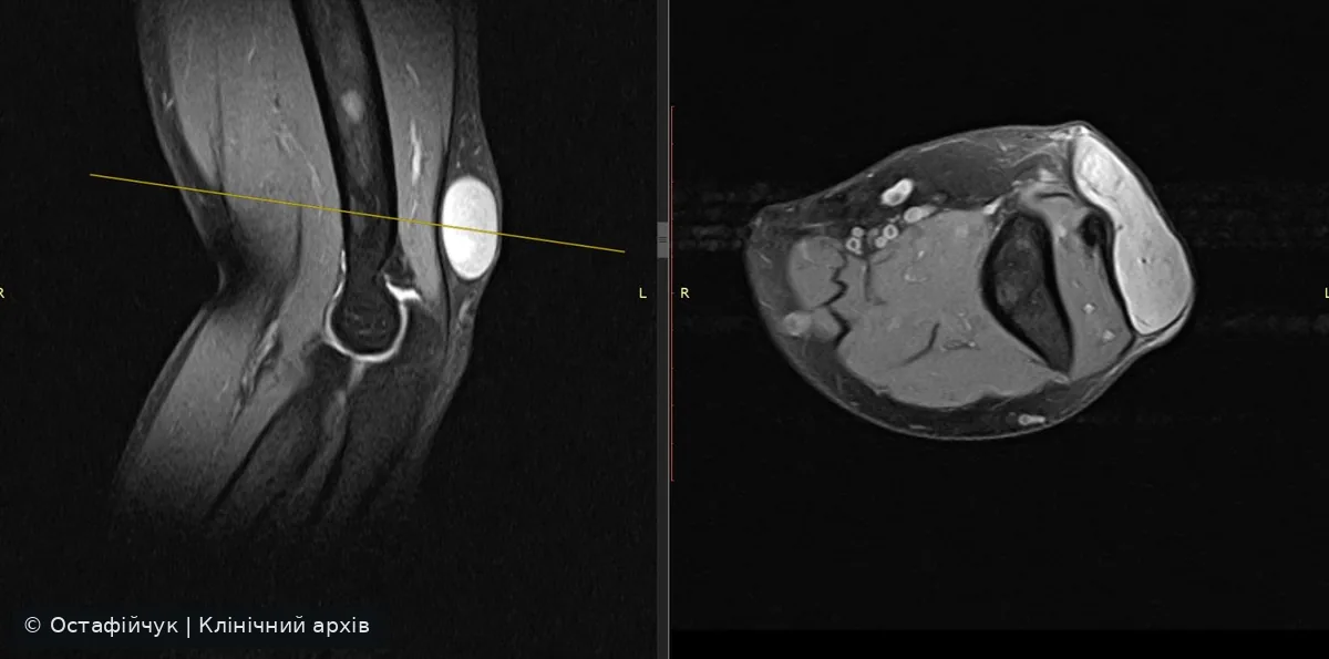
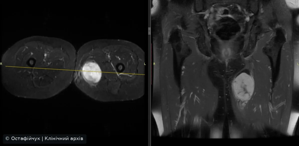
Avoiding diagnostic overreach
- Do not derive histological type from CT or MRI signal alone.
- Correlate radiographs, full-bone MRI, clinical context and older studies.
- Do not biopsy before the specialist team agrees a safe tract.
- Not every lesion requires biopsy: confidently benign findings may follow other pathways.
Biopsy planningTests needed before sampling →Chondrosarcoma caseConfirmed humeral tumour →Pubic GCT caseDiagnosis and resection planning →
Clinical sources
- UK guidelines for the management of bone sarcomas, 2025
- ESMO-EURACAN-GENTURIS-ERN PaedCan, Bone Sarcoma CPG
- NCI Osteosarcoma Treatment (PDQ)
- NCI Ewing Sarcoma Treatment (PDQ)
Clinical images and videos from the author’s archive. Patient identifiers have been removed. Diagnoses are stated only where supported by source records. This information does not replace an individual multidisciplinary decision.