Bone tumour biopsy: why it is planned before surgery
A bone tumour biopsy must provide adequate tissue for diagnosis, and its trajectory is planned so that the biopsy tract can be removed during definitive surgery when required.
Imaging before biopsy
Initial radiographs show bone architecture, while MRI or CT defines intramedullary extent, soft-tissue extension and relationships with important structures. Imaging helps target viable tissue and avoids a route that would be difficult to remove later.
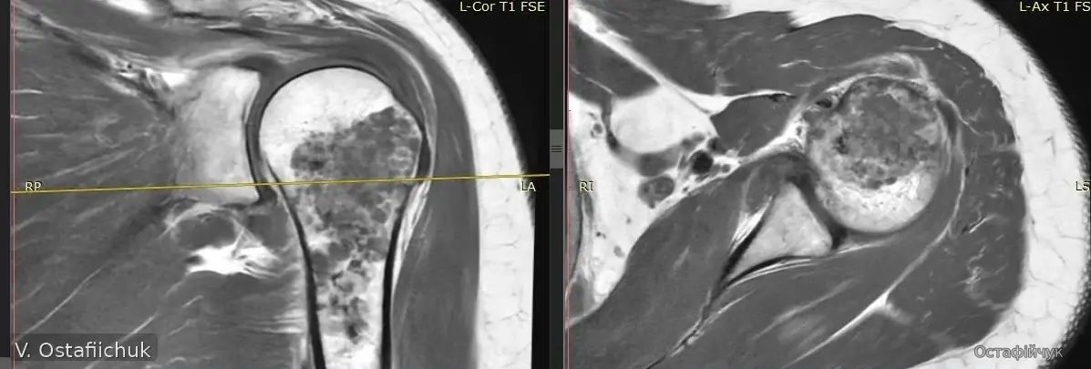
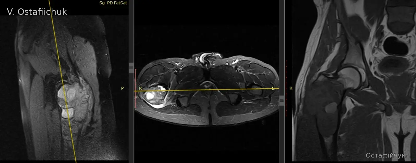
Clinical material from the author's archive is published with confirmed consent. Images are de-identified and illustrate principles; they do not replace assessment of an individual case.
Why the biopsy channel matters
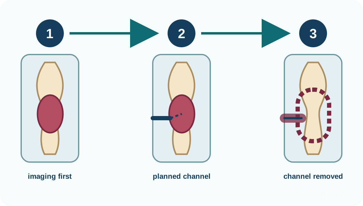
When a primary malignant bone tumour is suspected, the biopsy tract is planned as a potentially contaminated area that should, whenever possible, be included in the future resection. It should not cross several compartments, approach the neurovascular bundle unnecessarily or conflict with the definitive surgical approach.
Percutaneous or open biopsy
Core needle biopsy
Fluoroscopy, ultrasound or CT guidance can obtain multiple tissue cores from a representative area. Tissue quantity and processing requirements should be agreed in advance.
Open / incisional biopsy
This may be needed after a non-diagnostic percutaneous biopsy or when adequate tissue cannot be obtained safely otherwise. The incision is planned within the future surgical approach; in an extremity it is usually longitudinal and positioned so that it can be removed during definitive surgery.
Where the biopsy should be performed
Ideally it is planned by the team responsible for definitive treatment or in direct coordination with that team. The pathologist needs the clinical and radiological context because diagnosis depends on correlating morphology, imaging and clinical course.
Important: not every bone tumour is malignant. Benign, intermediate and malignant lesions follow different pathways, so treatment should not be prescribed from the appearance of one scan alone.