Skin tumour biopsy
A well-planned biopsy provides tissue for an accurate diagnosis without compromising the next stage of treatment.
Start with the clinical question
The method is selected according to what pathology must answer, where the lesion is, whether it can be removed in full and how a definitive operation might be performed.
Four principal methods
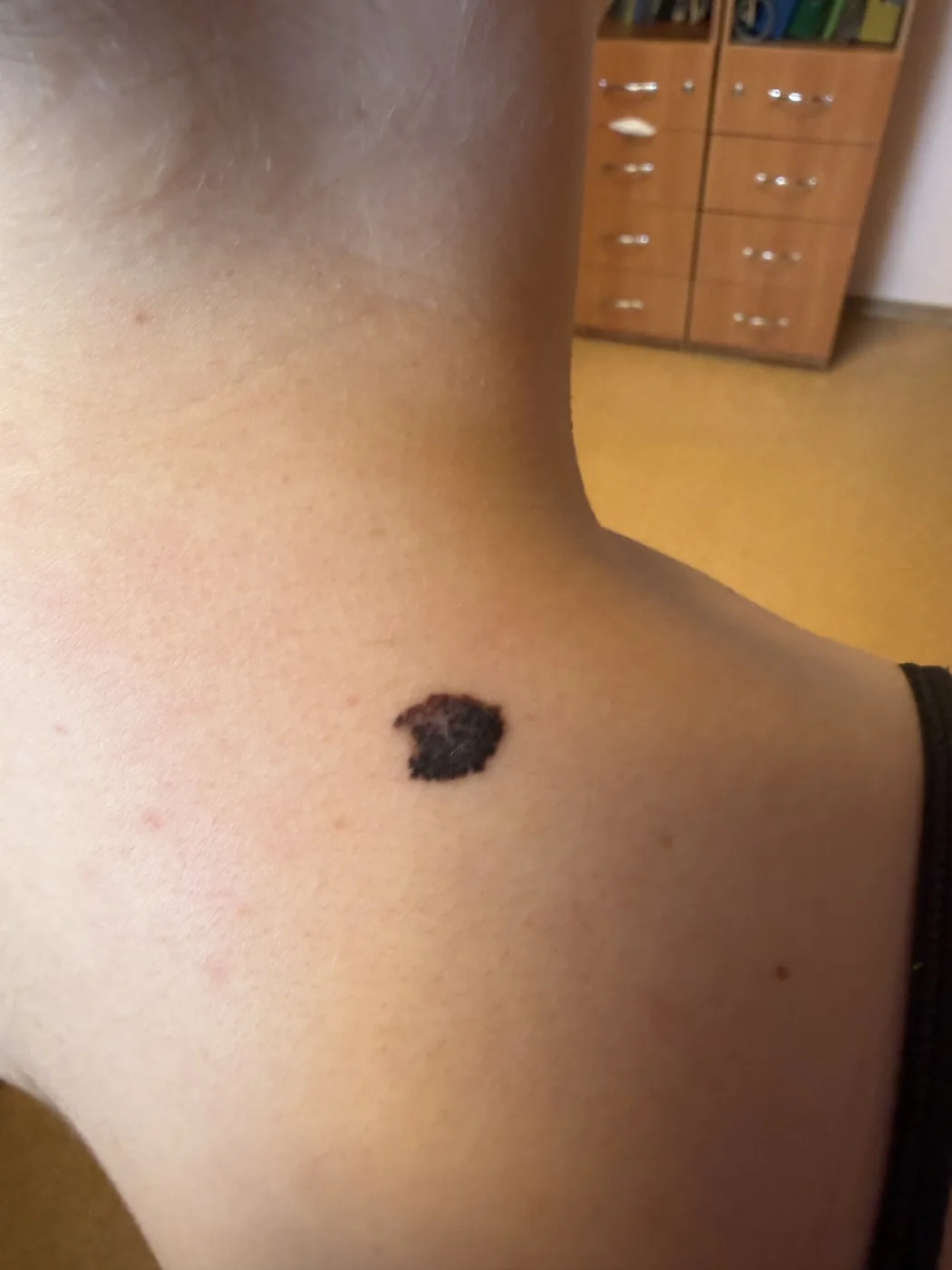
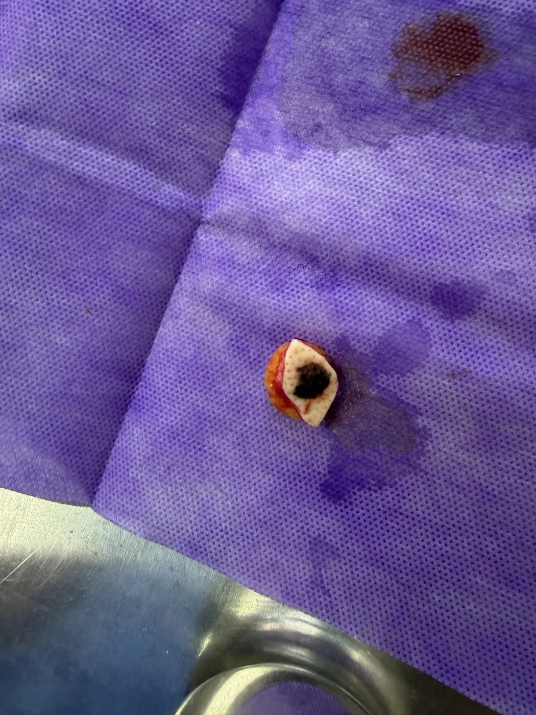
Removal of the entire visible lesion for diagnosis. Often preferred for a small suspicious pigmented lesion when anatomically feasible.
Read more →Punch biopsyA cylindrical full-thickness sample. Useful for partial sampling, but not a universal method when melanoma is suspected.
Read more →Incisional biopsyA representative part of a large or difficult lesion is sampled so the future operation can incorporate the biopsy track.
When it is used ↓Shave / saucerizationA tangential or deeper saucerization sample. Depth must answer the clinical question; a superficial shave may prevent accurate Breslow measurement.
Limitations ↓Skin biopsy: how the method is selected
This Ukrainian-language video shows why punch, excisional and other biopsy approaches are chosen according to the lesion, the clinical question and what pathology needs to answer.
Clinical footage: the video contains footage of a medical procedure.
Incisional biopsy
It may be used when complete diagnostic removal would create an excessive defect or when a tumour is large. The site and direction are planned around the definitive oncological operation.
Shave and saucerization
A superficial shave is appropriate only for selected superficial lesions. Deeper saucerization can provide a more complete sample, but a suspected invasive melanoma should not be transected at its deepest point if accurate Breslow measurement is to be preserved.
What pathology needs
- an intact and appropriately oriented specimen;
- the exact anatomical site;
- the clinical suspicion and any previous procedure;
- margin orientation where relevant.
What happens after a biopsy
After tissue is sampled, it undergoes laboratory processing, slide preparation and morphological assessment. Standard staining may be sufficient in some cases; others require additional sections, immunohistochemistry or expert slide review. There is therefore no single turnaround time that applies to every biopsy.
How long does a histology result take?
In this Ukrainian-language video I explain why speed matters, but an accurate morphological diagnosis matters more than an arbitrary universal deadline.
Clinical images from the author's archive, published with confirmed consent.