Excisional skin biopsy
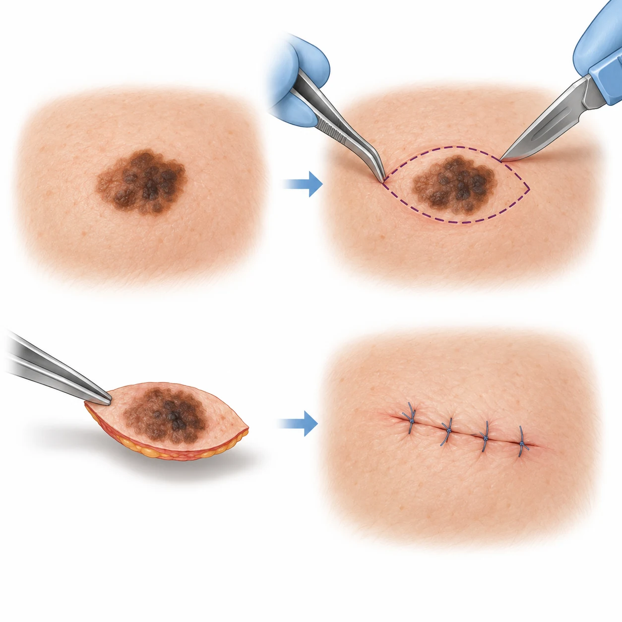
Complete removal of the visible lesion with a small diagnostic margin — to obtain an accurate pathology report and plan the next step.
What “excisional” means
In an excisional biopsy, the lesion is removed in full as a diagnostic procedure. The specimen is sent to a pathologist, who confirms the tumour type and reports features that determine further management.
When it may be appropriate
It is considered when the lesion can be removed safely in full, while preserving specimen orientation and without compromising future treatment. The choice depends on size, site, clinical suspicion and the question pathology must answer.
What happens
- Assessment, marking and local anaesthesia.
- Removal of the lesion as one oriented specimen.
- Wound closure and transfer of tissue for histology.
- Review of the report and, where needed, planning of the next procedure.
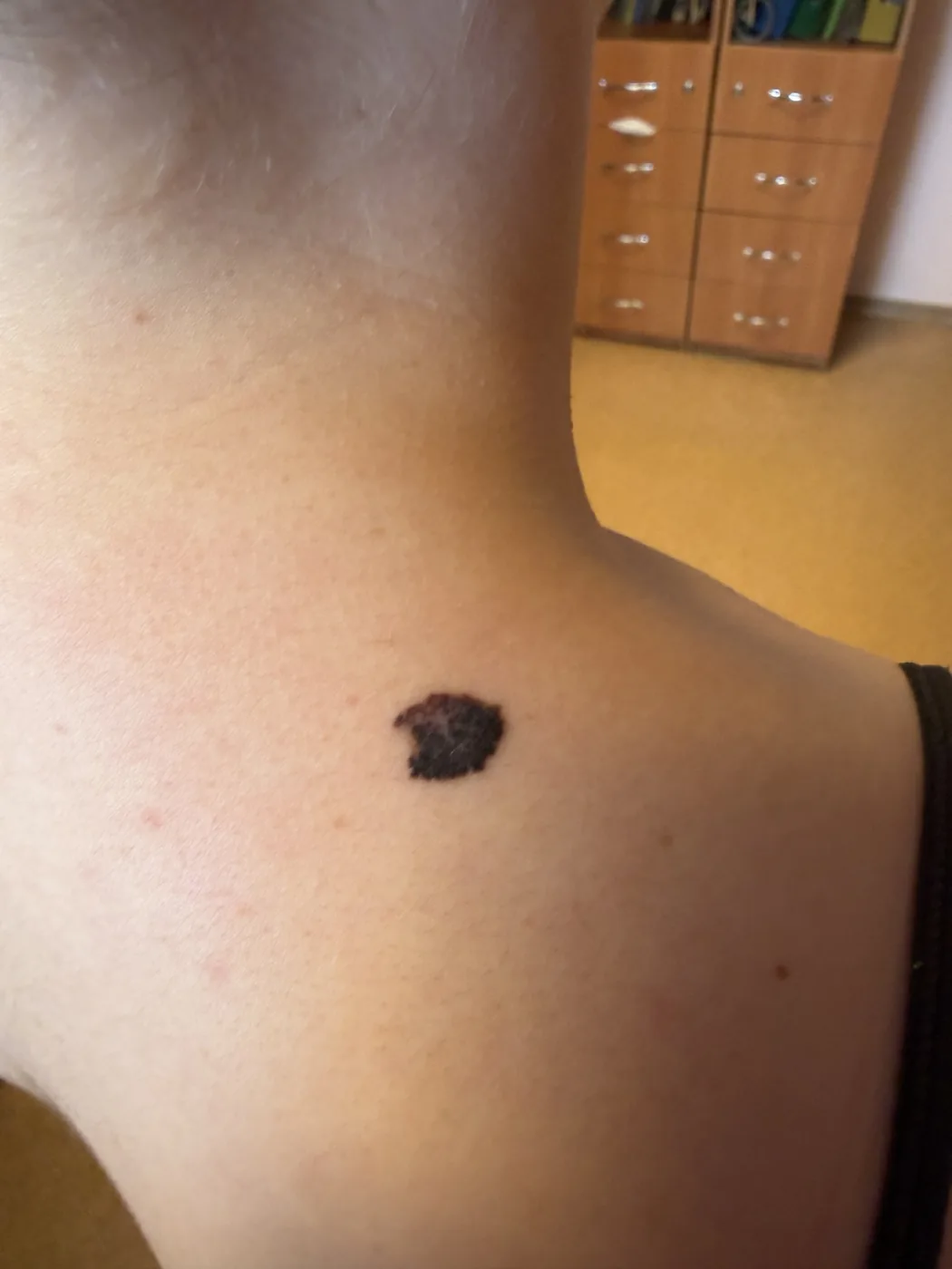
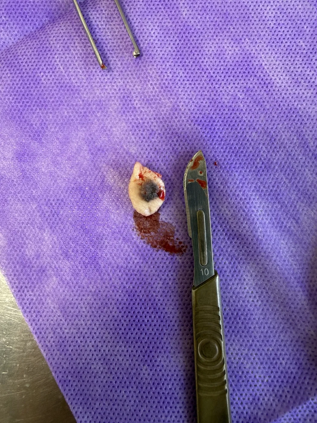
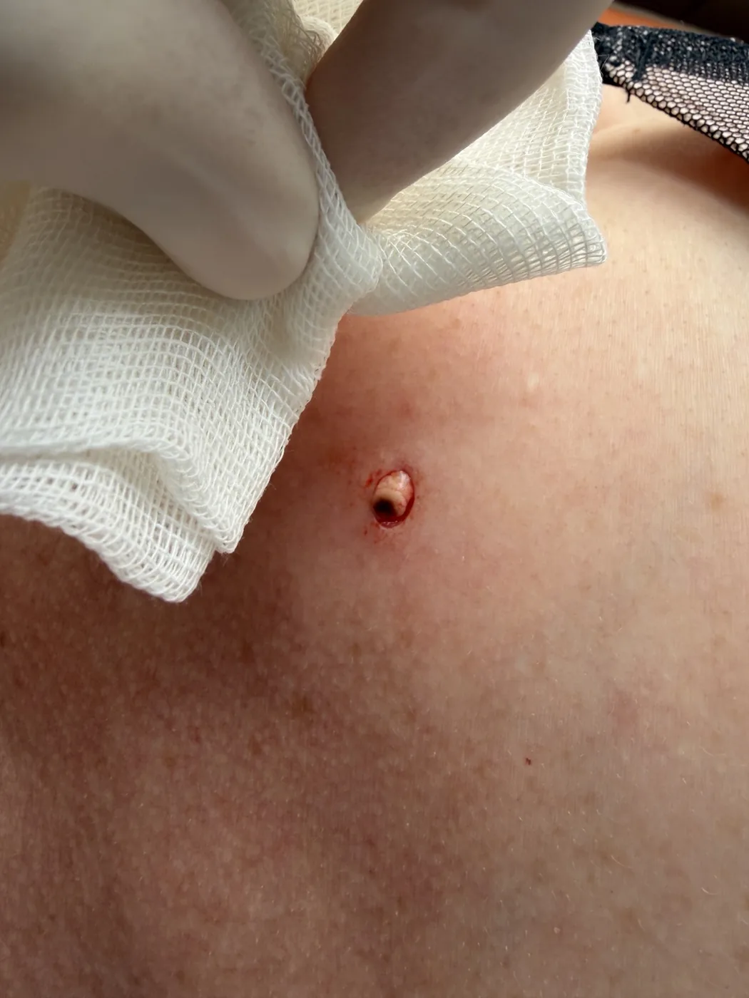
Separate clinical examples
The three images below are independent educational examples. They are not a single patient sequence.
Short clinical clip
No sound or autoplay. Only a short procedural step is shown.
What matters in the report
Further decisions depend on more than the diagnostic label. In melanoma, Breslow thickness and ulceration are among the critical features; in other tumours, histological type, risk factors and specimen margins may guide the plan.
Clinical material from the author's archive, published with confirmed consent. Final management follows clinical assessment and pathology.
This educational material does not replace clinical examination, a complete pathology report or an individual treatment plan.