Rare malignant skin tumours
Rare skin cancers may require a different biopsy, pathology panel, margin strategy and staging pathway. Precise subtype matters.
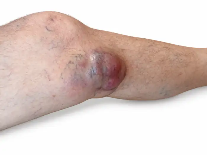
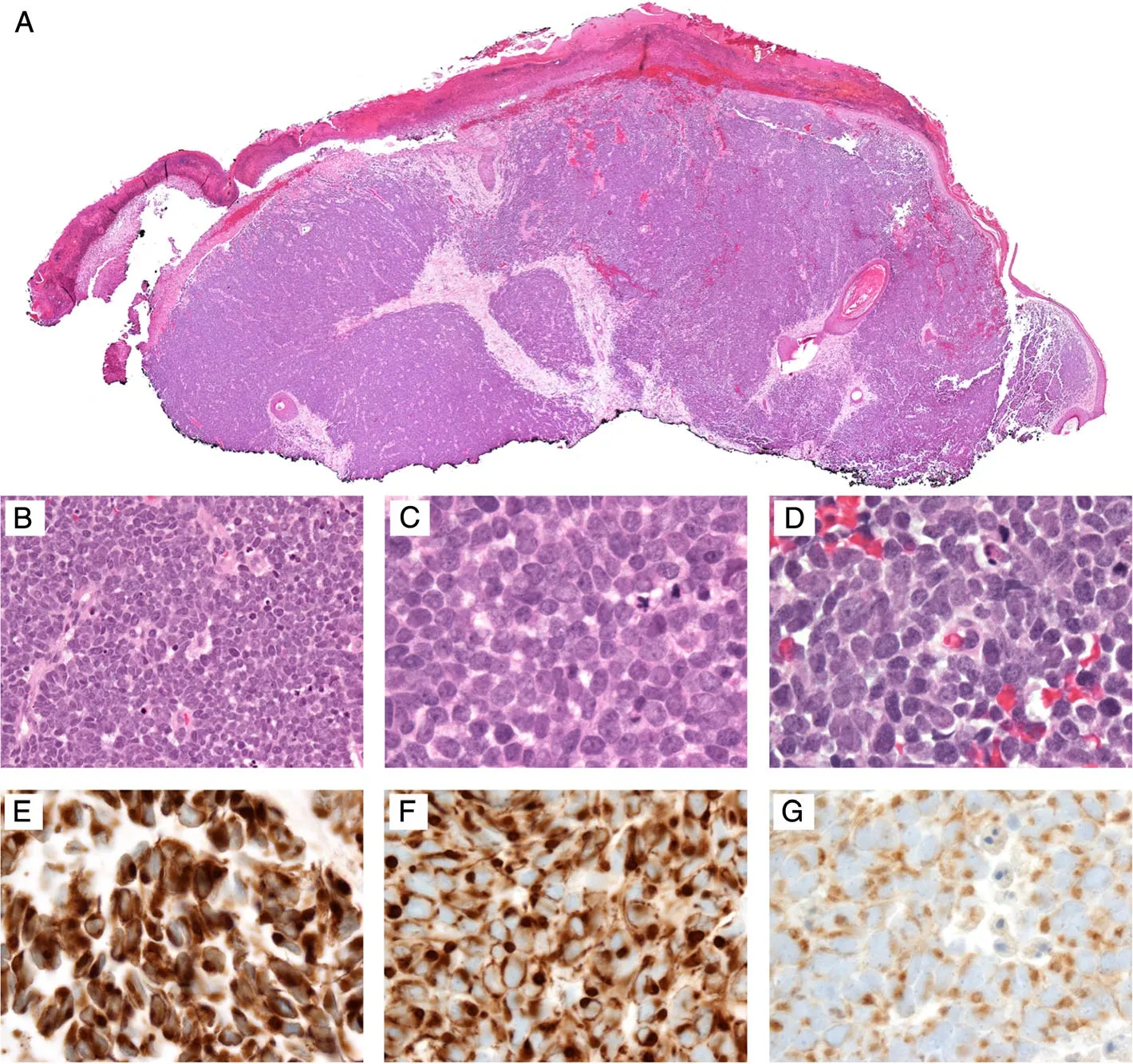
Merkel cell carcinoma
A rapidly growing red-violet nodule may look nonspecific, but MCC has a distinct SLNB, radiotherapy and systemic-treatment pathway.
Clinical: author case, histologically confirmed, published with permission. IHC panel: Nguyen AH et al., CC BY 4.0.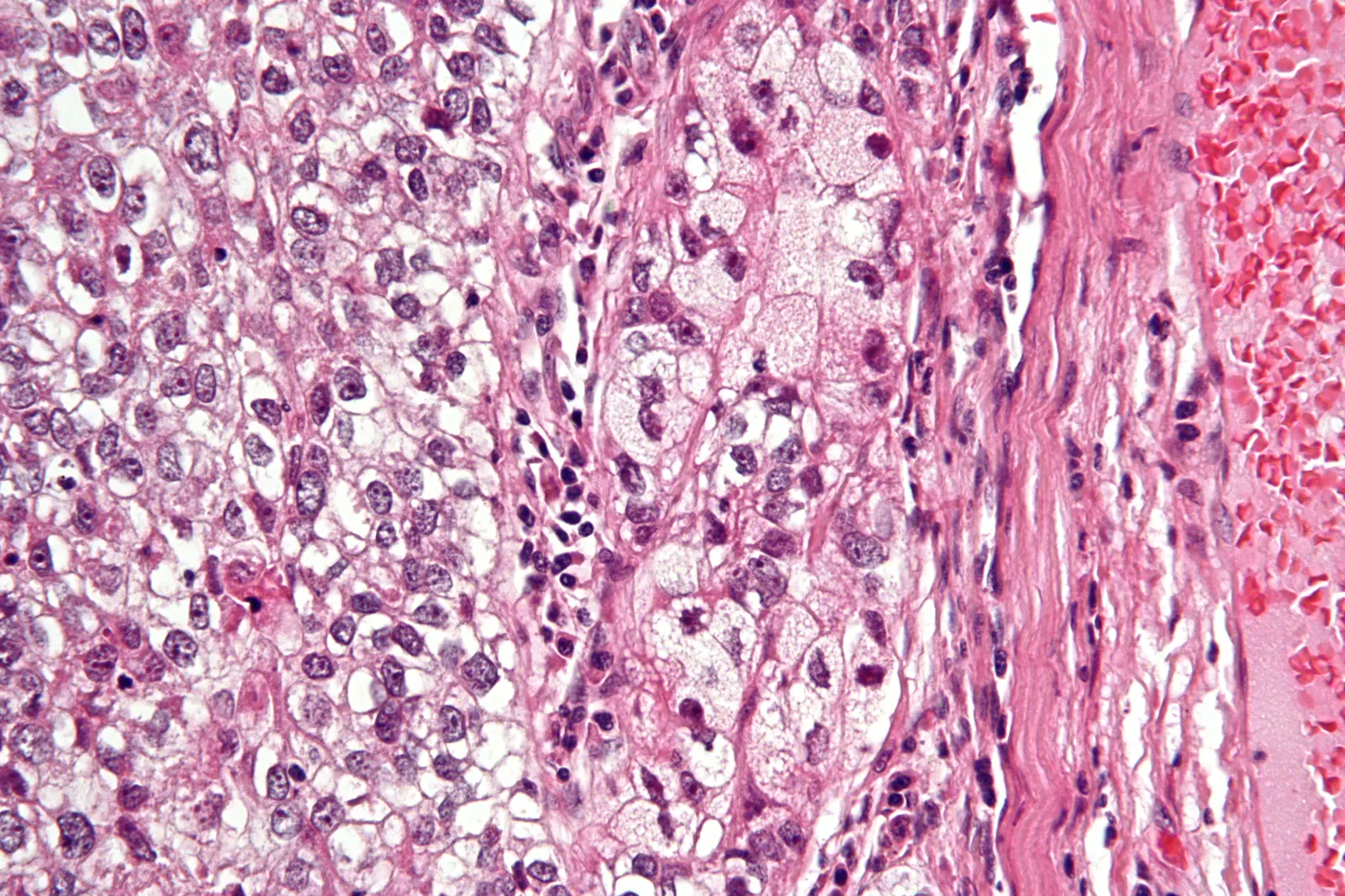
Sebaceous carcinoma
Particularly important in eyelid tumours. MMR IHC can be a useful screening tool, but an abnormal result alone does not diagnose Lynch syndrome.
Nephron / Wikimedia Commons, CC BY-SA 3.0.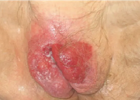
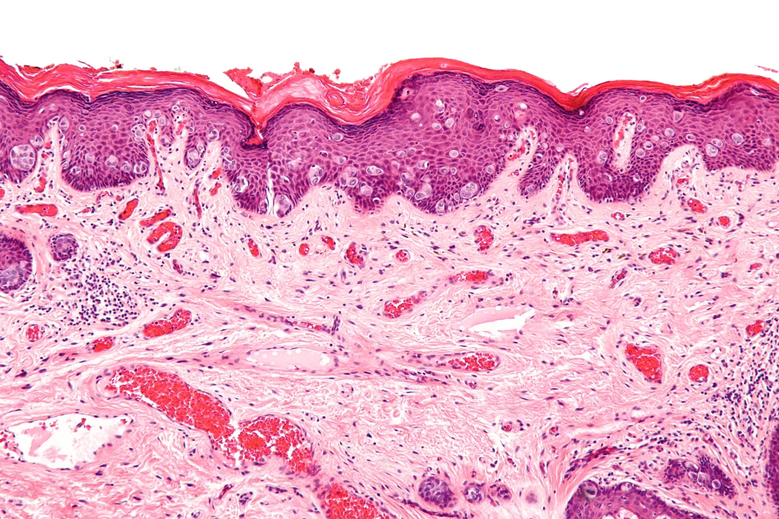
Extramammary Paget disease
A persistent eczema-like lesion at an apocrine-bearing site deserves biopsy and distinction between primary cutaneous disease and secondary spread from an internal carcinoma.
Clinical: Londero AP et al., CC BY-SA 4.0. Histology: Nephron, CC BY-SA 3.0.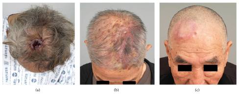
Cutaneous angiosarcoma
An enlarging bruise-like, violaceous or infiltrated scalp lesion deserves early biopsy because the visible border can underestimate disease extent.
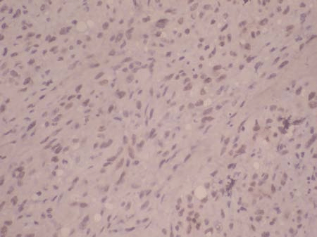
Clinical: Choi JH et al. / Wikimedia Commons, CC BY 4.0. Histology: Deenadayal D et al., CC BY 4.0.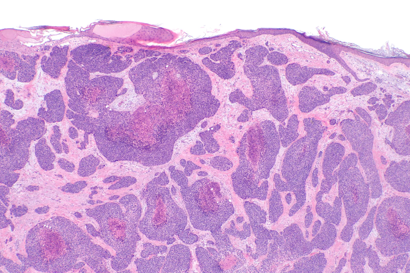
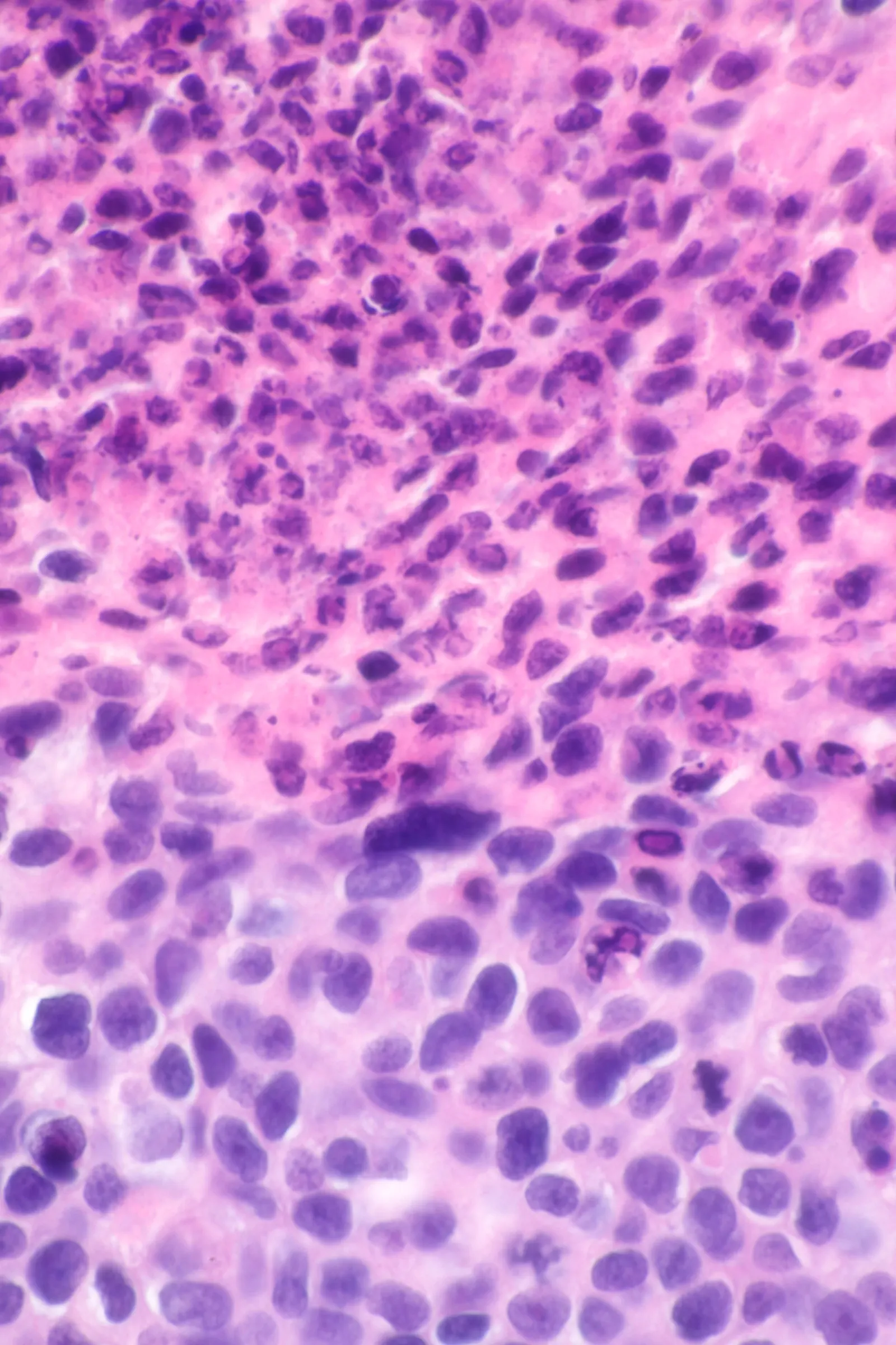
Porocarcinoma and adnexal carcinomas
These tumours may mimic cSCC or a benign adnexal lesion clinically; IHC and molecular features may be needed to resolve the diagnosis.
Histology, low and high magnification: Nephron / Wikimedia Commons, CC BY-SA 4.0.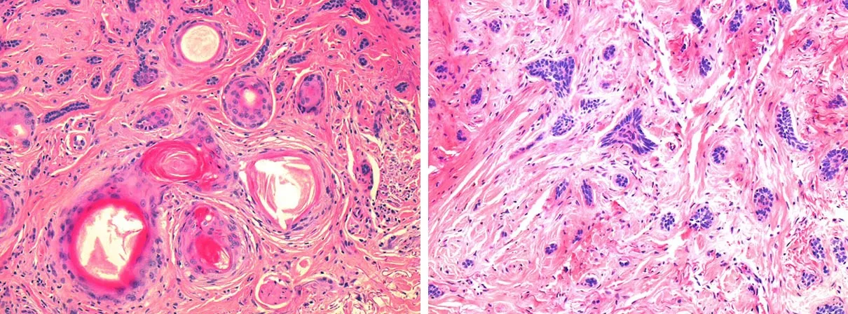
Microcystic adnexal carcinoma
Clinically indolent-appearing disease may extend much deeper than expected. A superficial biopsy can underestimate its true extent.
Mike Inskip, Jill Magee / Wikimedia Commons, CC BY 4.0.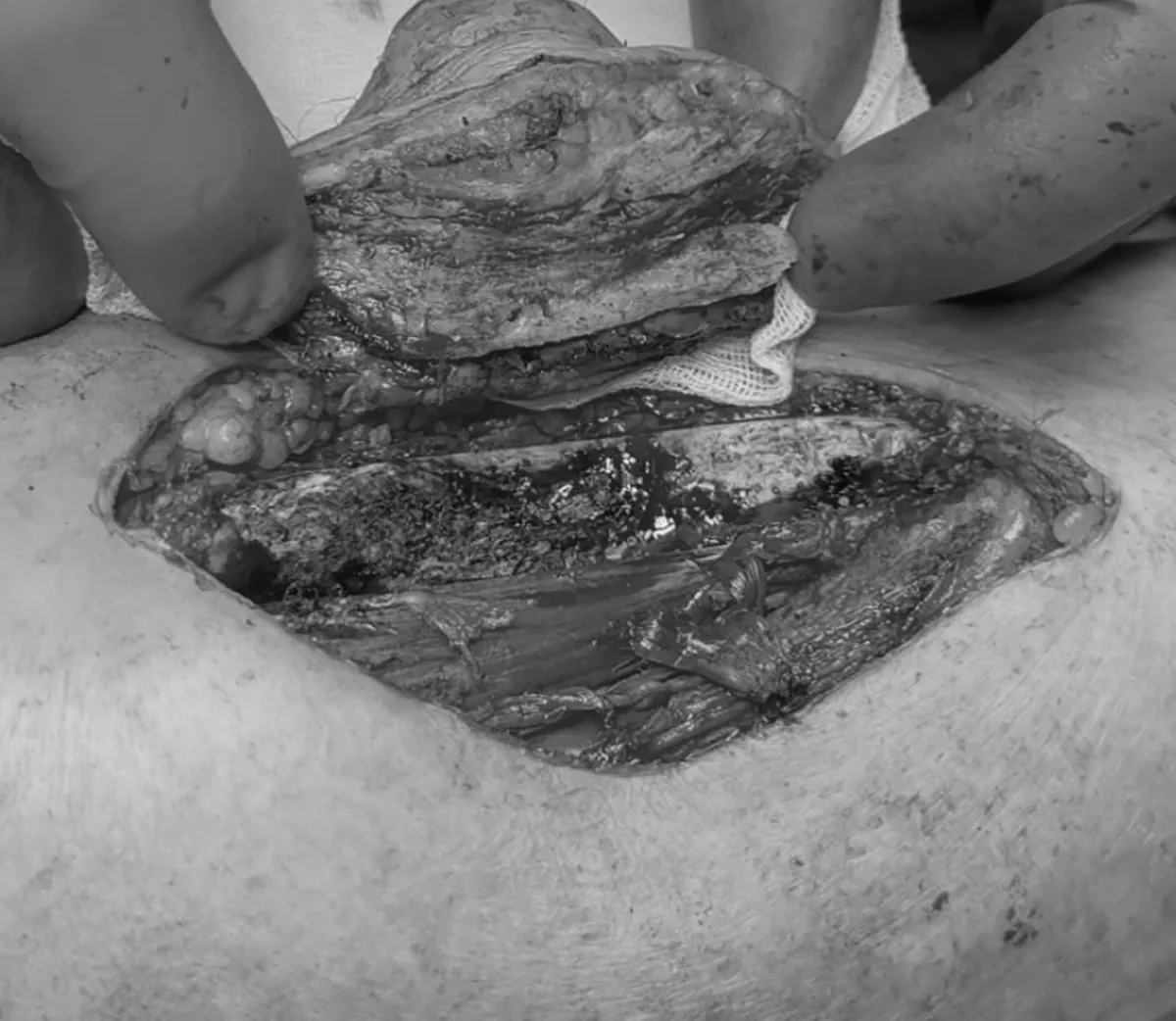
Dermatofibrosarcoma protuberans
A detailed DFSP pathway is already available in the sarcoma cluster, including surgery, molecular confirmation and reconstruction.
Author clinical material.Clinical clues by site and appearance
IHC and molecular clues - a differential-diagnosis map
A dot-like CK20 pattern supports MCC, but diagnosis depends on a coherent panel and exclusion of small-cell lung carcinoma, melanoma and lymphoma.
MCC pathway →In the appropriate clinical context, tumour MMR IHC can select patients for genetic assessment, but an abnormal tumour profile alone does not prove Lynch/Muir-Torre syndrome.
Sebaceous carcinoma →The IHC panel should follow the anatomic site and differential diagnosis to distinguish primary cutaneous EMPD from secondary epidermotropic spread of an internal carcinoma.
EMPD →Morphology is paired with vascular markers; the visible skin border may underestimate true disease extent, so pathology and imaging should be interpreted together.
Angiosarcoma →In difficult cases, molecular and IHC features can help separate porocarcinoma from cSCC, but they are not mandatory universal tests for every tumour.
Adnexal carcinomas →Deep infiltrative cords and perineural spread may lie beyond a superficial specimen, so a shallow biopsy can materially underestimate the tumour.
MAC →How not to lose a rare tumour inside the generic label “skin cancer”
The main risk with rare cutaneous malignancies is applying a BCC or cSCC pathway simply because the tumour arose in skin. Biopsy depth, IHC, molecular confirmation, margin strategy, nodal staging and systemic therapy can all differ substantially by entity.
The sample must be sufficient to assess architecture and depth, not only superficial cytology.
Morphology plus IHC, with molecular confirmation or review when it changes classification.
Imaging, SLNB and screening for an internal primary are not the same across all rare skin cancers.
Surgery, margin control, reconstruction, radiotherapy and systemic treatment should fit the exact entity.
What else belongs to the adnexal-tumour spectrum
Other rare entities include digital papillary adenocarcinoma, primary cutaneous adenoid cystic carcinoma, mucinous carcinoma, apocrine carcinoma and additional sweat-gland malignancies. For these tumours, expert pathology and complete margin assessment are particularly important.
Rare-skin evidence map 2024-2026
Recent updates extend beyond tumour names. Sebaceous carcinoma evidence is refining MMR screening, EMPD guidance now integrates underlying-malignancy work-up and HER2-directed systemic therapy, porocarcinoma literature highlights YAP1/NUT in difficult diagnosis, and new population data for MAC reinforce the importance of deep margin control.
Sweat-gland carcinoma review 2026 · MMR-IHC meta-analysis 2026 · EMPD guideline 2025 · Angiosarcoma guideline 2025 · Porocarcinoma review 2024 · MAC population study 2026.