Dermatofibrosarcoma protuberans (DFSP)
A rare, usually slow-growing skin tumour with infiltrative local growth, where accurate diagnosis and margin control are central.
What it is and how it may appear
Dermatofibrosarcoma protuberans (DFSP) is a skin tumour that often grows slowly. It may look like a firm patch, plaque or nodule that gradually enlarges without pain. Appearance alone cannot confirm the diagnosis.
Why biopsy matters
A planned biopsy establishes the diagnosis under the microscope and helps plan treatment. For a large or deep tumour, the team decides whether imaging is needed to assess its extent.
DFSP staging: why it differs
Unlike cutaneous melanoma, DFSP has no universally used dedicated I–IV stage grouping. Clinicians describe its extent and behaviour: whether it is localized or advanced, superficial or deep, primary or recurrent, and whether fascia, muscle, regional sites or distant organs are involved. Pathology also looks for fibrosarcomatous transformation.
| Clinical feature | What it means | Why it matters |
|---|---|---|
| Localized DFSP | Tumour remains in the skin and adjacent tissue, without detected spread. | The team maps the true extent and whether complete removal is possible. |
| Deep local extension | Tumour reaches fascia, muscle or deeper structures. | This may change imaging, excision and reconstruction planning. |
| Recurrent DFSP | Tumour returns after earlier treatment. | The prior scar and possible hidden extension matter. |
| Fibrosarcomatous transformation (FS-DFSP) | Pathology identifies a more aggressive component. | Multidisciplinary review and follow-up planning are important. |
| Regional spread | Nearby nodes or other regional sites are involved. | The team clarifies extent and an individual plan. |
| Distant metastatic disease | Tumour is found in distant organs. | This is advanced disease requiring multidisciplinary assessment. |
Framework: NCCN DFSP, version 1.2025; European interdisciplinary guideline, update 2024 (published 2025). This table describes clinical extent, not AJCC stage groups.
Why the visible border may be misleading
DFSP can extend as narrow projections through skin and subcutaneous tissue beyond what can be seen or felt. The operation is therefore planned beyond the visible outline alone.
Excision, margins and reconstruction
Depending on site, size and the available pathology assessment, treatment may involve wide excision or micrographically controlled removal. Resection margins are the peripheral and deep edges of the specimen; pathology checks whether tumour cells reach them. A positive margin calls for an individual discussion of further management. Closure with a flap or another method follows definition of the adequate oncological extent.
One clinical case sequence
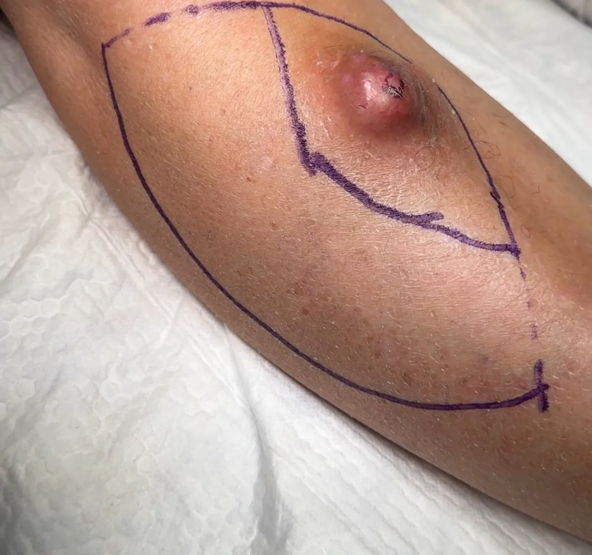
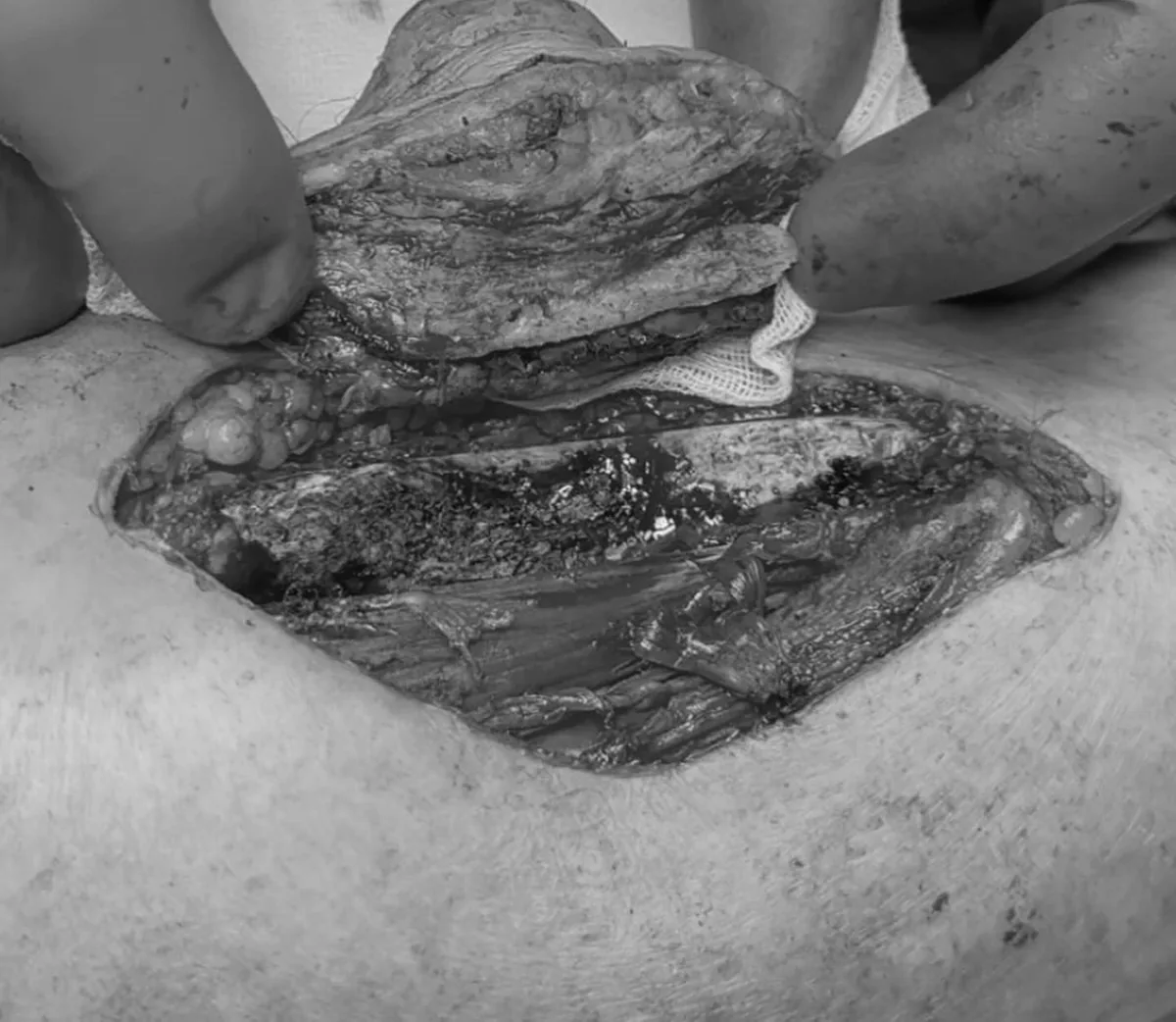
The post-excision defect and oriented specimen.
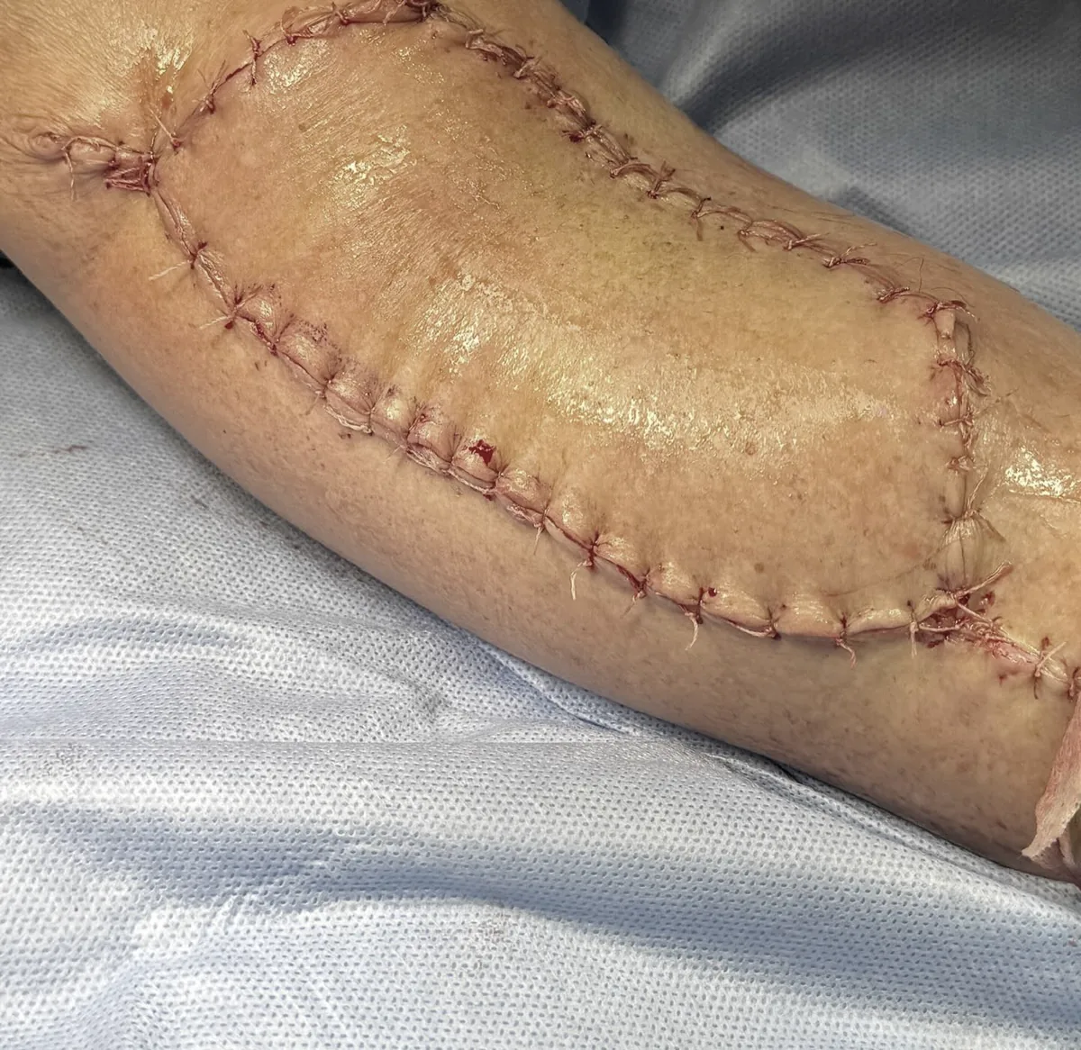
Follow-up after treatment
Visits assess the operative site and any new skin changes. The schedule depends on final pathology, margin status and the clinical situation.
One clinical case from the author's archive, published with confirmed consent. Social overlays were removed and the graphic step was desaturated.
This educational material does not replace clinical examination, a complete pathology report or an individual treatment plan.