Reconstruction after skin cancer removal
First, adequate cancer removal. Then, restoration of form and function with the most appropriate reconstructive method.
The oncological step determines reconstruction
Closure must not dictate an inadequate excision. Margins and the likely defect are planned together, but definitive reconstruction follows adequate tumour control.
The reconstructive ladder
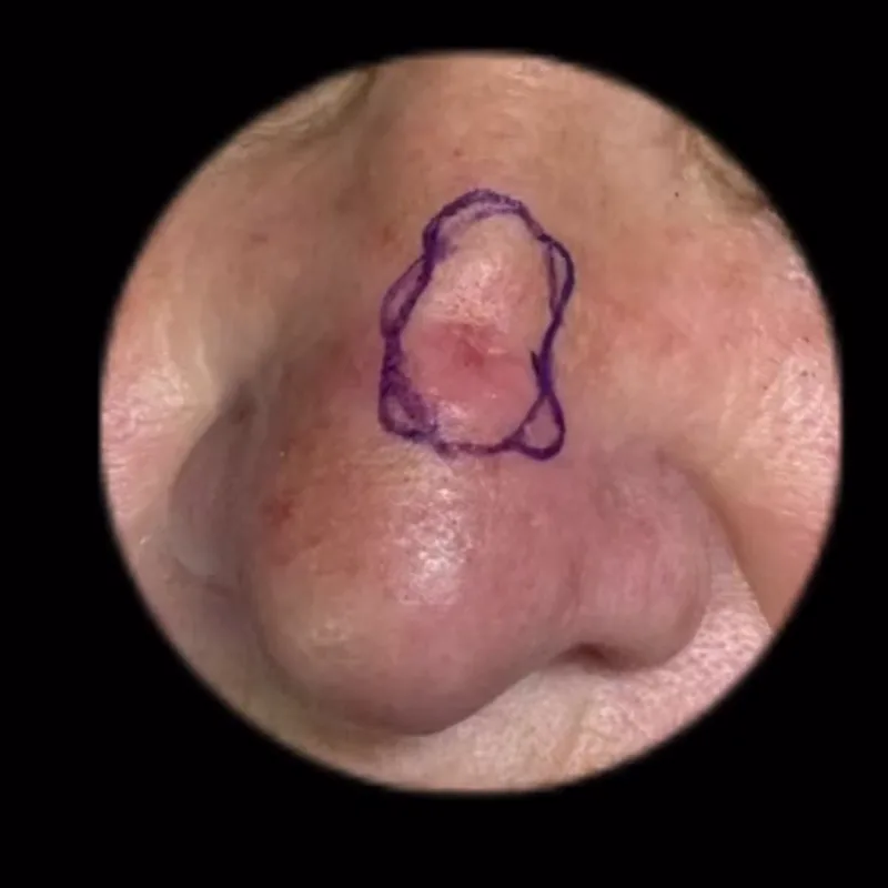
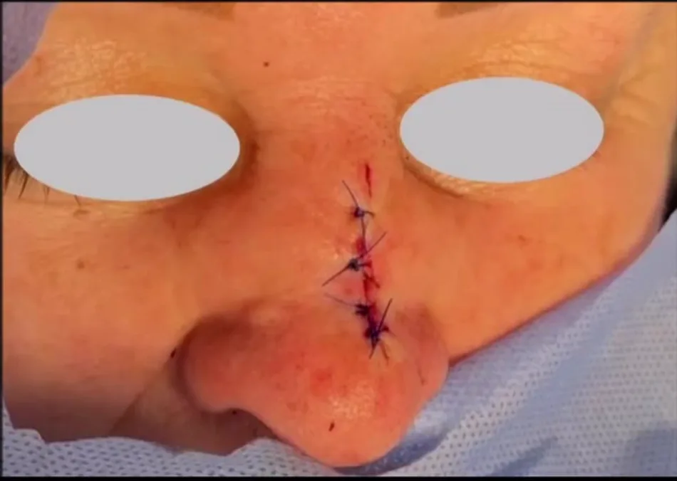
Nose - direct closure after margin assessment
A separate nasal basal cell carcinoma case: marking and immediate direct closure. These images do not show the full microscopic assessment or a late cosmetic result.
Face and periocular area — one clinical case
After defining the oncological defect, local flap repair considers nasal and eyelid form and function. Both images are from one patient; immediate closure is not the final scar appearance.
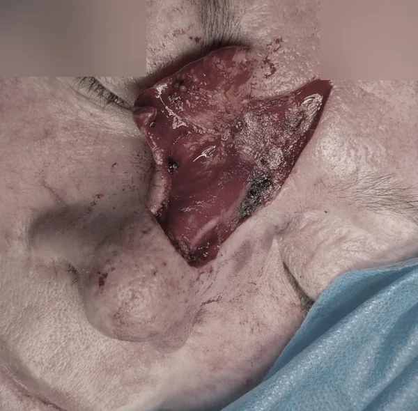
After tumour removal: a defined defect in an anatomically complex area.
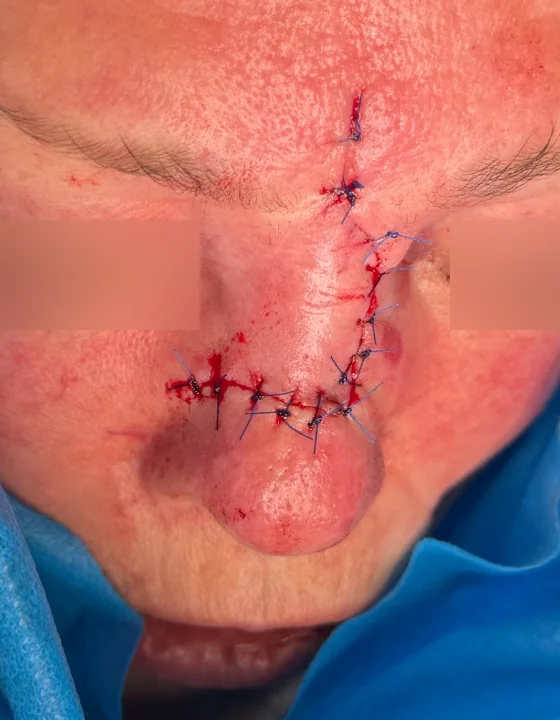
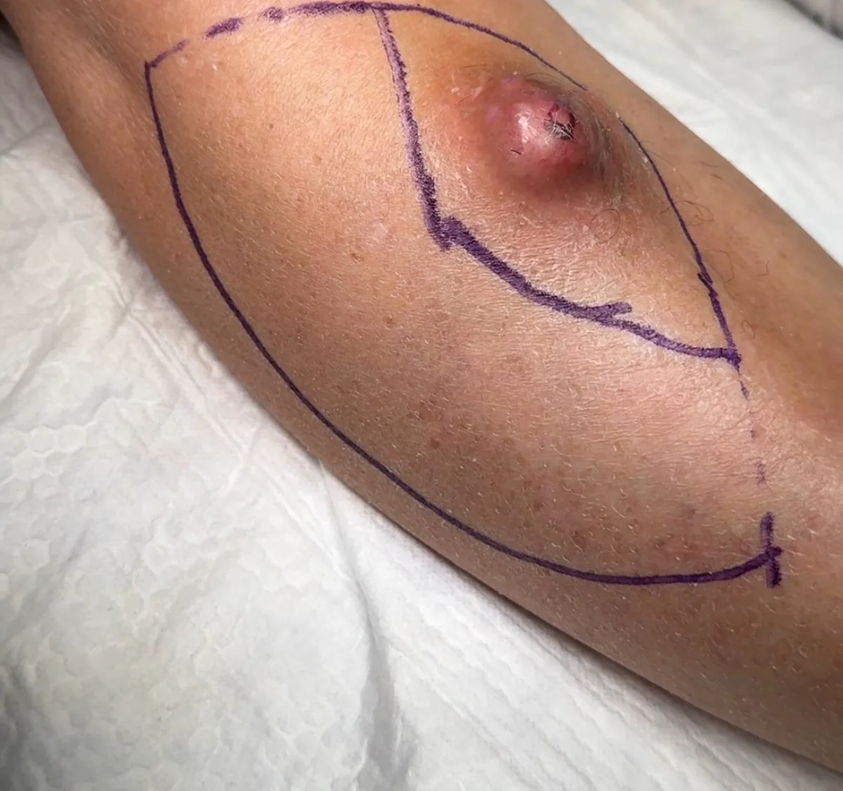
A Keystone flap for a larger defect
These three images show one clinical case: planning, the post-excision defect and immediate local-flap closure. The middle image is graphic.
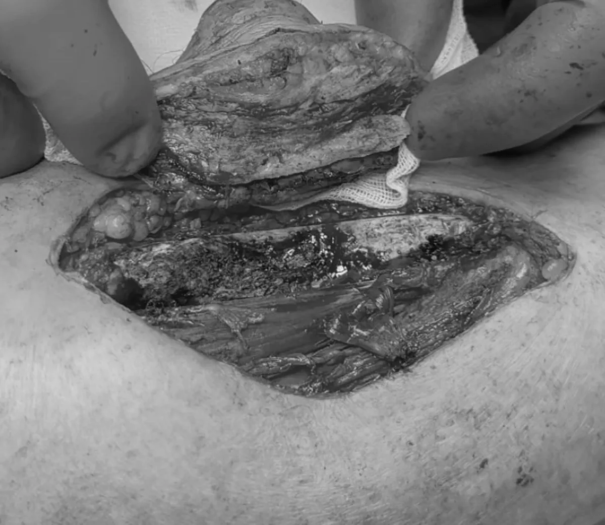
After excision: the defect and oriented specimen.
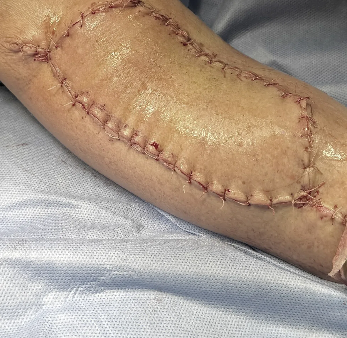
What is assessed afterwards
Tissue viability, healing, function, final margins and the need for further treatment. A final cosmetic result cannot be judged from an immediate postoperative image.
Clinical material from the author's archive, published with confirmed consent. Social overlays were removed and the graphic frame was desaturated.
This educational material does not replace clinical examination, a complete pathology report or an individual treatment plan.