Mohs micrographic surgery
Staged tumour removal with mapping and microscopic examination of margins — for selected tumours in difficult anatomical sites.
The principle
The tumour is removed in thin stages. Each layer is oriented, mapped and examined microscopically; if tumour remains in a specific area, further tissue is removed precisely from that location.
When it may be discussed
Common reasons include high-risk BCC or SCC, recurrent tumours, indistinct clinical borders and critical facial sites where preserving healthy tissue is especially valuable. Not every patient needs this technique.
Clinical example 1 - nasal tumour
One case: after biopsy, marking, an excision stage and immediate closure. The images do not themselves prove clear microscopic margins; that requires examination of oriented tissue.
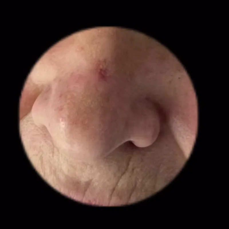
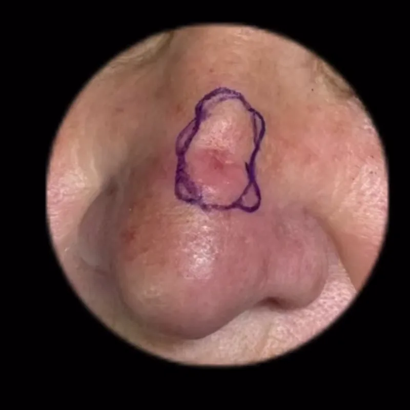
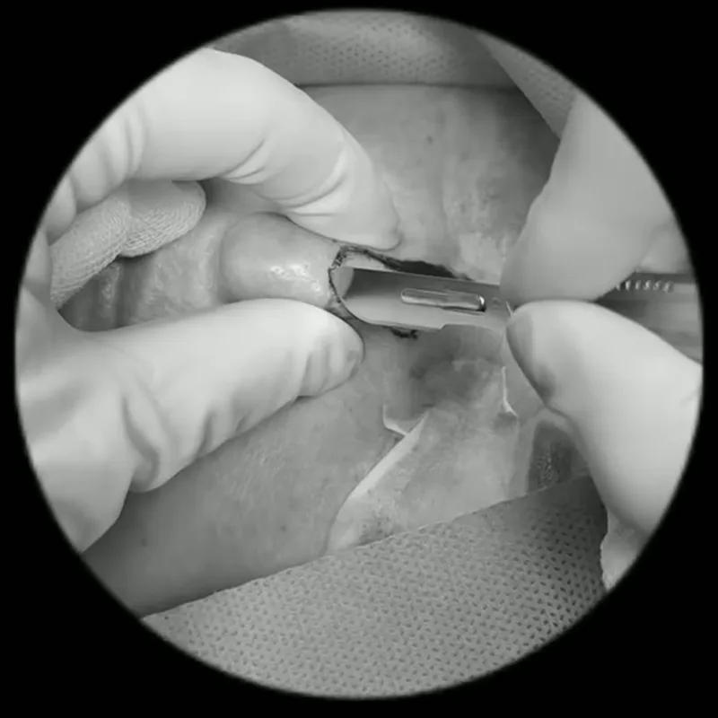
An excision step. The oriented specimen is assessed for margins; this image does not show microscopy.
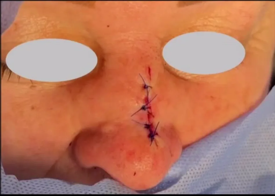
Clinical example 2 — defect and local flap
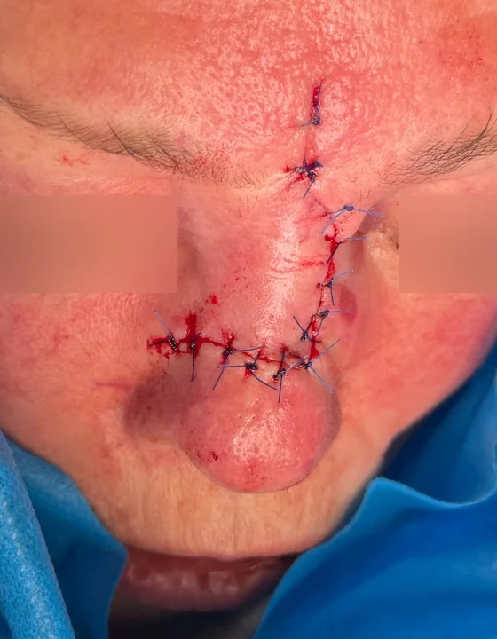
These two images show one patient: the defined defect and immediate closure. The photographs alone do not document microscopic margin assessment.
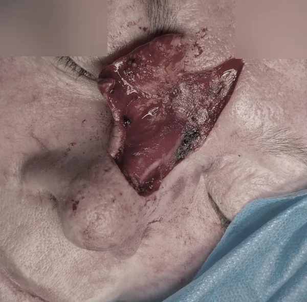
After tumour removal: a defined defect in an anatomically complex area.
Separate short surgical clip
A different clinical case: this brief silent step does not continue either example above.
00:00–00:10 — a short stage of removal. The frame was cropped to the operative field, converted to greyscale and stripped of audio.
When reconstruction is performed
The defect is closed after adequate margin control. Direct closure, a local flap, a graft or staged reconstruction may be selected according to site, size and functional structures.
Material from the author's archive, published with confirmed consent. The video does not replace individual planning.
This educational material does not replace clinical examination, a complete pathology report or an individual treatment plan.