Basal cell carcinoma (BCC)
The most common skin cancer usually grows locally, but in difficult sites it can damage important structures and require precise margin control.
Clinical TNM, pathological groups, site distinctions and staging limitations.
When to seek assessment
A non-healing ulcer, pearly or reddish nodule, repeated bleeding, crusting or slow enlargement - particularly on sun-exposed skin - should be assessed.
Diagnosis
Dermoscopy helps evaluate a lesion, but histology establishes the diagnosis. The biopsy method depends on size, site and the anticipated treatment.
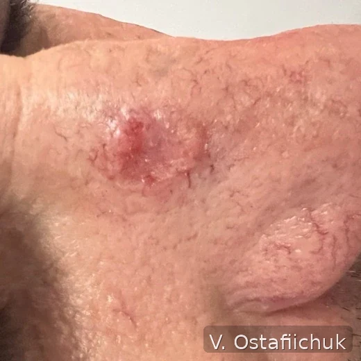
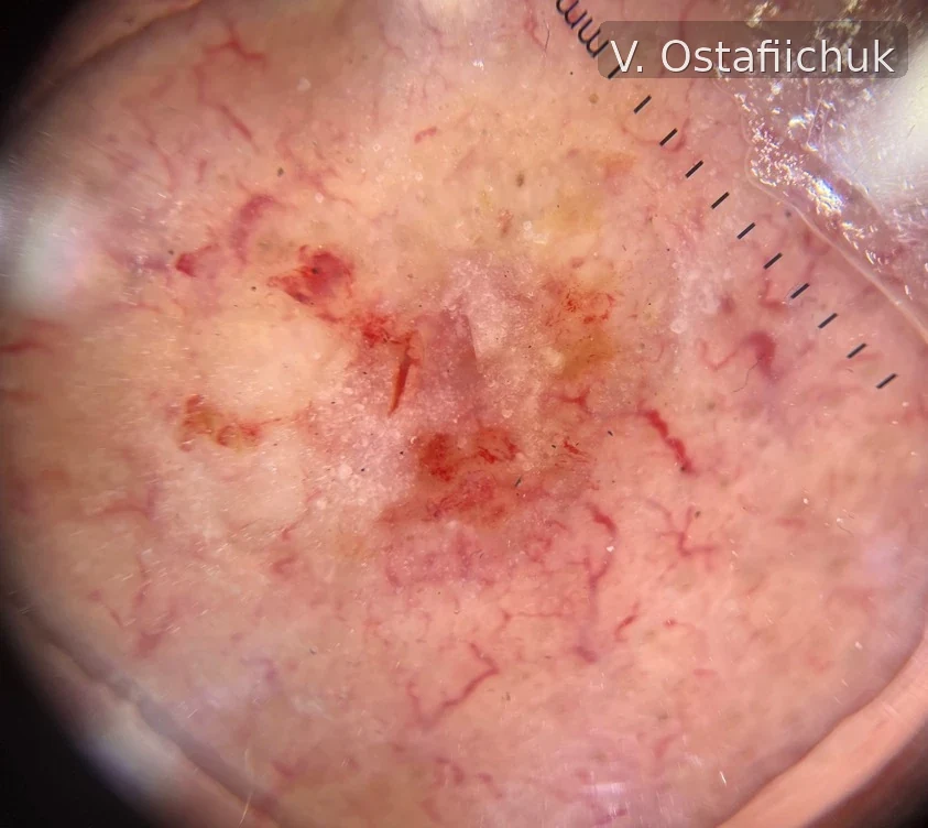
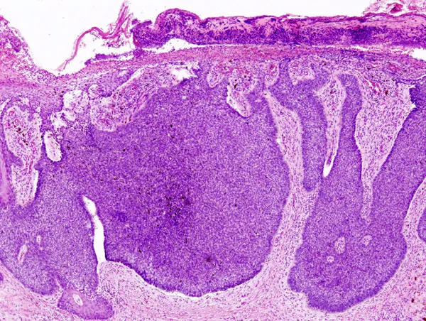
Clinical appearance, dermoscopy and histology
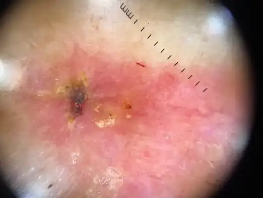
Author clinic + dermoscopy example
The same BCC is useful to assess at several levels: clinical appearance shows site and visible borders, dermoscopy adds structural information, while final classification remains histologic.
How treatment is selected
Histological subtype, size, border definition, primary or recurrent status, site and patient factors all matter. Surgery is the main treatment for most operable BCCs. Staged micrographic margin control may be appropriate for high-risk disease or critical facial sites.
Not all BCC is the same
Lower risk
A typical primary tumour in a favourable site where complete excision is straightforward.
Higher risk
Critical site, recurrence, poorly defined borders, aggressive histology or other local-risk features.
Difficult-to-treat
Disease where standard excision may create unacceptable functional harm or cannot reliably control the tumour.
Margin-controlled surgery, including Mohs, is particularly useful in selected high-risk and anatomically critical BCC. Hedgehog inhibitors and anti-PD-1 treatment belong to advanced unresectable/metastatic scenarios, not routine local BCC.
Recurrence after prior treatment: a separate clinical scenario
Recurrent BCC should not be assessed as simply “another similar spot”; previous treatment and the current clinical findings are part of the problem. The author presentation contains a clinical example labelled as solid basal cell carcinoma of the skin - recurrence after radiotherapy.
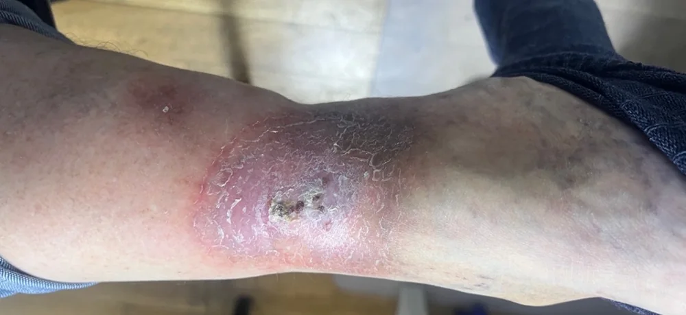
What this case demonstrates: previous treatment history is part of the clinical problem. Planning for recurrence depends not only on appearance but also on prior treatment, anatomical site, pathology and the feasibility of reliable margin control.
Periocular planning and closure: separate examples
These archive images illustrate separate clinical examples, not one patient sequence.
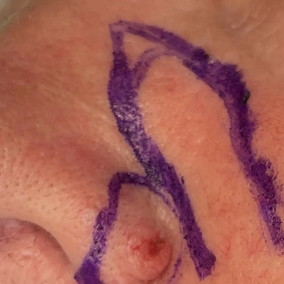
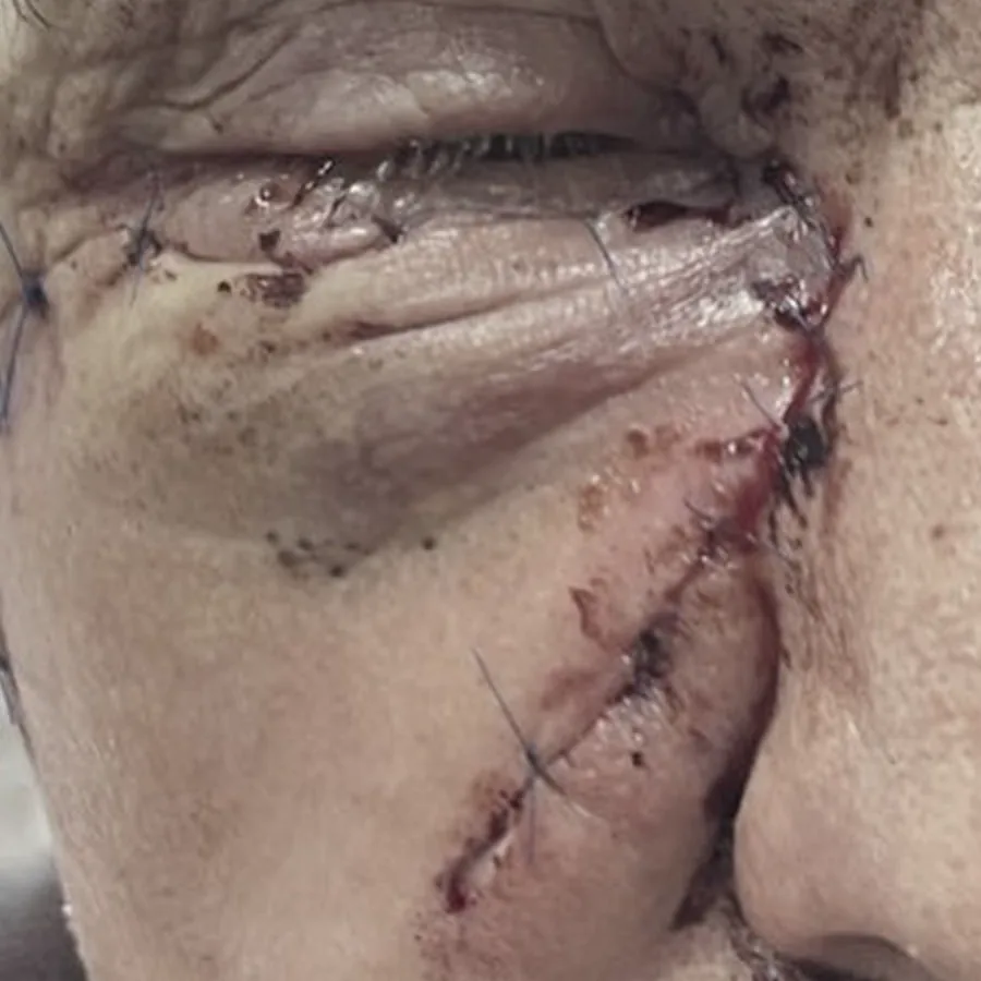
Nose: Mohs + reconstruction - an author clinical pathway
On the nose, even a small BCC can be difficult because tissue is limited and tumour control must be balanced with alar shape, nostril patency and reconstruction. The sequence below uses author clinical material to show the logic from biopsy to closure.
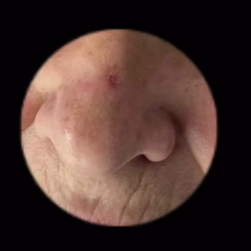
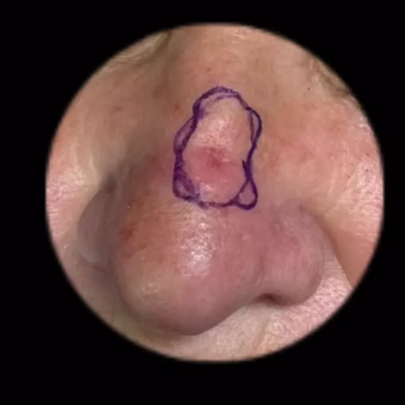
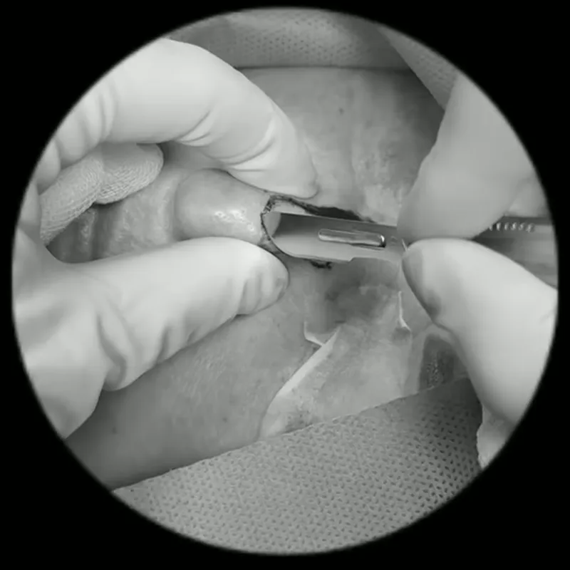
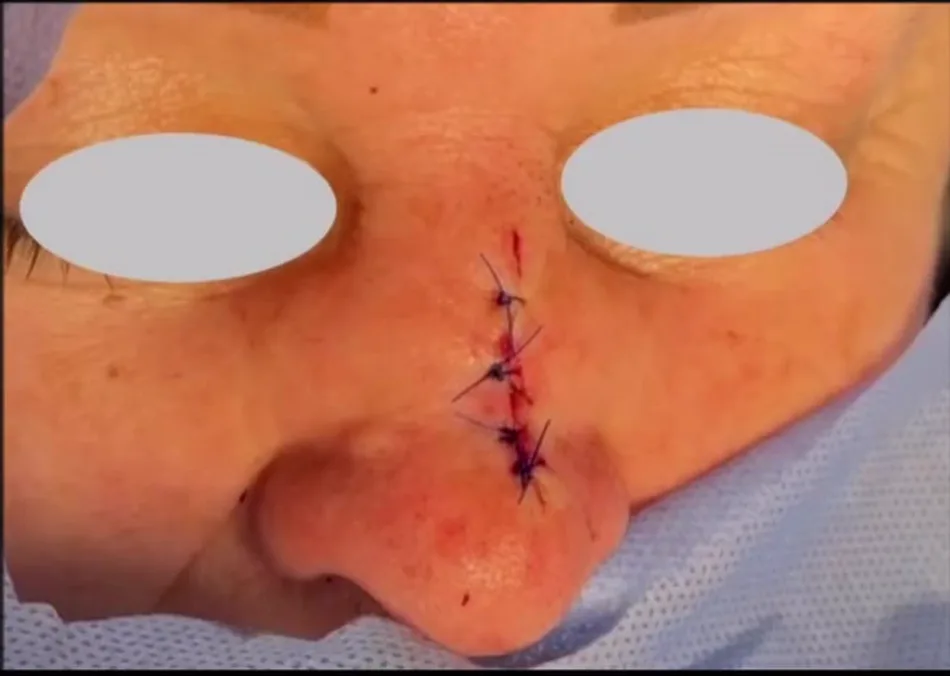
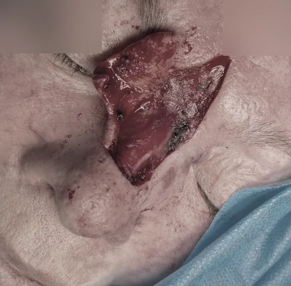
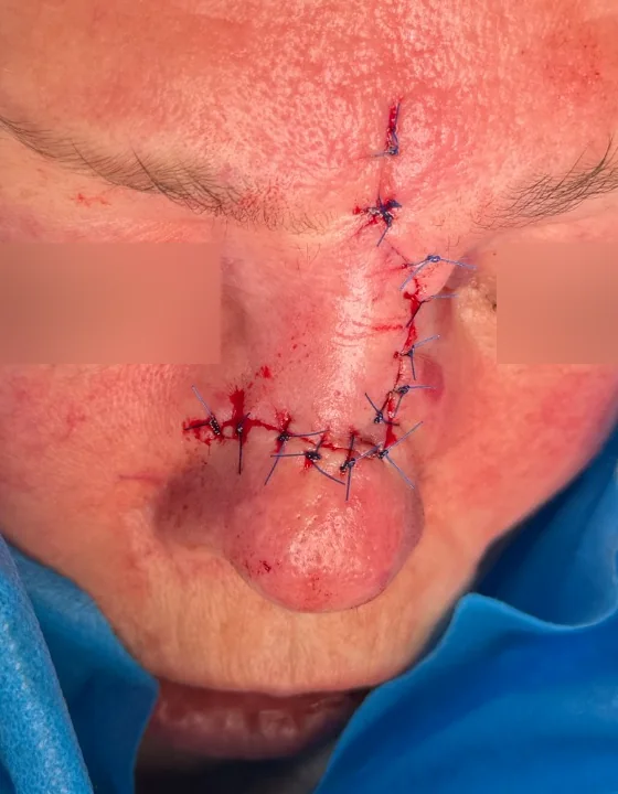
Detailed nasal BCC + Mohs case · When Mohs is considered · Reconstruction after skin cancer.
AJCC 8 for cutaneous carcinoma of the head and neck
This AJCC 8 system applies to cutaneous squamous cell and basal cell carcinoma of the head and neck; it is not a universal staging system for every skin site. Eyelid carcinoma has a separate AJCC 8 chapter and TNM system.
| Stage | TNM / grade | What it means |
|---|---|---|
| Tis | Tis | Carcinoma in situ. |
| T1 | T1 | Tumour ≤2 cm. |
| T2 | T2 | Tumour >2-4 cm. |
| T3 | T3 | Tumour >4 cm, or minor bone erosion, deep invasion or defined perineural invasion. |
| T4a / T4b | T4a / T4b | T4a is gross cortical bone or marrow invasion; T4b is skull-base or skull-base foramen invasion. |
| N0 / N1 | N0 / N1 | N0: no regional nodal metastasis. N1: one ipsilateral node ≤3 cm without extranodal extension. |
| N2 / N3 | N2 / N3 | N2 covers specified single or multiple nodes up to 6 cm; N3 is a node >6 cm or clinically/pathologically significant extranodal extension. |
| M0 / M1 | M0 / M1 | M0: no distant metastasis; M1: distant metastasis. |
| 0 | Tis N0 M0 | Carcinoma in situ. |
| I | T1 N0 M0 | Tumour ≤2 cm without nodal or distant metastases. |
| II | T2 N0 M0 | Tumour >2-4 cm without nodal or distant metastases. |
| III | T1-T2 N1 M0; or T3 N0-N1 M0 | T3 without advanced nodal disease, or a smaller tumour with one small ipsilateral node and no extranodal extension. |
| IV | T1-T3 N2 M0; or T4 any N M0; or any T N3 M0; or any T/N M1 | More extensive nodal disease, bone/skull-base invasion or distant metastasis. |
Formal staging is rarely needed for BCC because distant spread is uncommon; local risk, margin assessment and feasibility of complete removal are usually more relevant.
Sources: AJCC Current Staging System 2026 - Cutaneous Carcinoma of the Head and Neck and Eyelid Carcinoma, 8th edition; NCI Skin Cancer Treatment PDQ.
Modern systemic layer
For locally advanced or metastatic BCC not adequately manageable with surgery or radiotherapy, Hedgehog inhibitors such as vismodegib or sonidegib are established options. Cemiplimab can be considered after HHI or when HHI is not appropriate.
2026 practical signal: HHI response may sometimes reduce the morbidity of subsequent surgery, but clinical or dermoscopic disappearance does not guarantee histologic eradication. In a 2026 real-world sonidegib series, complete pathologic clearance after 12 weeks was confirmed in only a subset of patients, supporting tissue confirmation before definitive local decisions.
European BCC guideline 2023 · Sonidegib pathology study 2026 · NHS England neoadjuvant vismodegib policy 2026.
The NHS England policy is jurisdiction-specific and should not be automatically treated as the standard of care in Ukraine.
Outlook and follow-up
Most BCCs are treated successfully, especially at an early stage. Follow-up includes the scar, the rest of the skin and ultraviolet protection.
Clinical images from the author's archive, published with confirmed consent.