Merkel cell carcinoma
A rare but aggressive neuroendocrine skin cancer. Biopsy, nodal staging and local/systemic treatment should be planned as one coordinated pathway.
Author clinical case
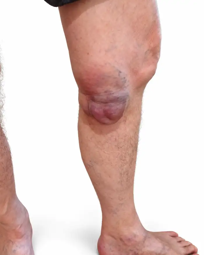
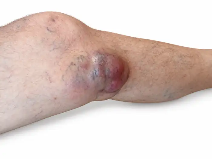
Below is the clinical appearance of histologically confirmed Merkel cell carcinoma in the knee region. The external appearance of MCC is nonspecific, so the photograph itself is not diagnostic - biopsy, morphology and immunohistochemistry remain decisive.
When to consider MCC
A new firm nodule growing over weeks to months, often on sun-exposed skin, needs biopsy. Clinical appearance alone is not diagnostic.
Pathology
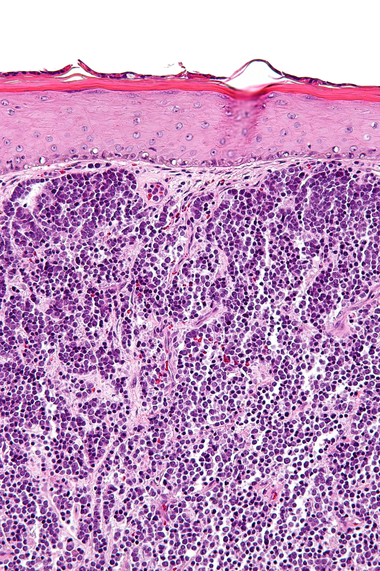
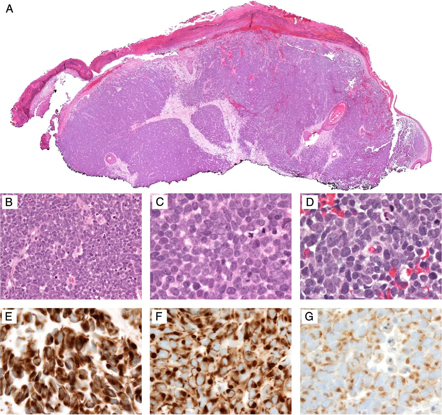
Morphology is supplemented by immunohistochemistry. A typical profile often includes CK20 with a characteristic perinuclear dot-like pattern, broad-spectrum cytokeratins and neuroendocrine markers. The diagnosis should rest on a coherent panel rather than one positive stain, helping distinguish MCC from metastatic small-cell lung carcinoma, melanoma, lymphoma and other small-round-cell mimics.
Nodes and sentinel-node biopsy
For clinically node-negative disease, sentinel lymph node biopsy is often an important staging step and, when indicated, is preferably planned before wide excision. Suspicious nodes require targeted confirmation.
Confirm MCC and perform appropriate IHC.
Assess nodes, image when appropriate and plan SLNB.
Surgery and radiotherapy according to stage and risk.
Immunotherapy for unresectable/metastatic disease; adjuvant approaches are individualized.
Staging before treatment selection
Once MCC is confirmed, staging should assess the primary tumour, regional lymph nodes and, when indicated, distant disease. A small primary lesion does not automatically imply low risk, so staging should be a deliberate step rather than an afterthought after surgery.
Primary tumour
Size, site, local extent and relevant pathological features.
Regional nodes
Clinical examination, targeted imaging when appropriate, and sentinel-node biopsy in suitable clinically N0 patients.
Distant disease
Imaging is selected according to stage, symptoms and risk; suspected metastases should be confirmed when that would change management.
Local treatment
For resectable localized MCC, oncologically planned excision is central. Radiotherapy often contributes substantially to local and regional control, but the field and timing depend on stage, pathology and nodal status.
If regional nodes are involved
Management differs between microscopic nodal disease detected by SLNB and clinically or radiologically evident nodal disease. Surgery, radiotherapy and systemic therapy may all be relevant, so nodal management is best planned within the same MDT pathway as the primary tumour.
Systemic therapy
For unresectable locoregional or metastatic MCC, PD-1/PD-L1 checkpoint inhibitors form the basis of modern systemic therapy. Drug selection and sequencing depend on prior therapy, access and the clinical situation. Cytotoxic chemotherapy can produce responses, but these are generally less durable and it is no longer the preferred first-line logic when immunotherapy is available.
Surveillance after treatment
Recurrence risk is highest in the first few years after treatment. Follow-up includes examination of the skin and surgical site, regional nodal assessment and imaging for appropriately selected higher-risk patients. Intensity should reflect stage, immune status and previous treatment.
ctDNA - a strong emerging biomarker, not a replacement for standard surveillance
Tumour-informed ctDNA has shown high sensitivity for active MCC and a strong association between a positive surveillance test and recurrence risk. In a 2026 multicentre cohort, ctDNA also outperformed MCPyV oncoprotein antibody testing for recurrence detection. This strengthens a risk-adapted surveillance role where testing is available, but does not make ctDNA mandatory for every patient or replace clinical review and appropriate imaging.
Prospective ctDNA study · ctDNA vs MCPyV antibodies, 2026 · Surveillance data 2026.
What is new
ctDNA is an emerging surveillance tool with promising prospective data, but it is not yet a universal standard for every patient.
Guideline anchors
SLNB, surgery, radiotherapy and systemic treatment should be selected according to stage in an expert multidisciplinary pathway.
Adjuvant immunotherapy in 2026: two phase III signals, but not yet a blanket standard
EA6174/STAMP: pembrolizumab did not reach statistical significance for the overall RFS primary analysis (p=0.10), but improved MCC-specific recurrence-free outcome (HR 0.66) and distant metastasis-free survival. ADAM: in node-positive MCC, avelumab produced an RFS HR of 0.61 (95% CI 0.32-1.16; p=0.132); the prespecified adjusted HR was 0.54 (p=0.069), while a non-prespecified relapse analysis also supported a treatment signal.
These trials move adjuvant checkpoint blockade much closer to routine MDT discussion, but do not support automatic treatment of every resected MCC patient without considering stage, radiotherapy, availability, regulatory status and individual risk.