Precancer and in-situ lesions
Actinic keratosis and Bowen disease
Not every rough patch is cancer, but persistent photodamage and carcinoma in situ need proper assessment, especially when a lesion thickens, bleeds or changes.
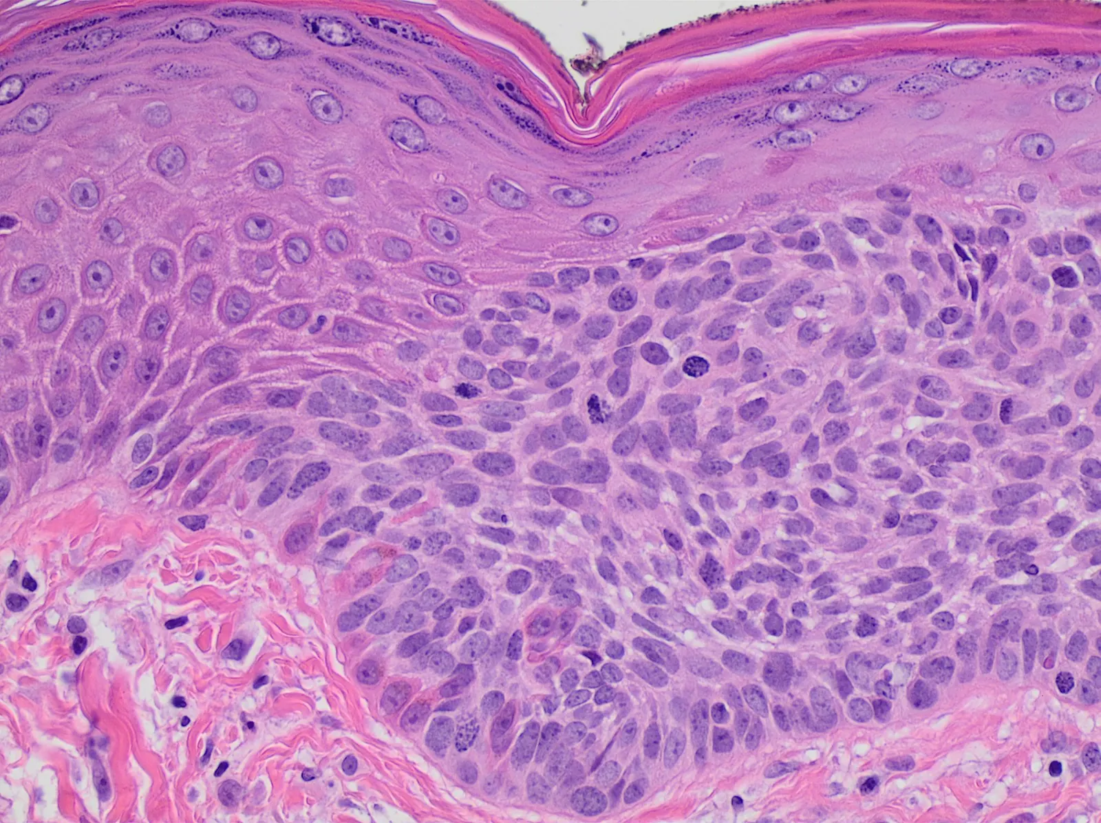
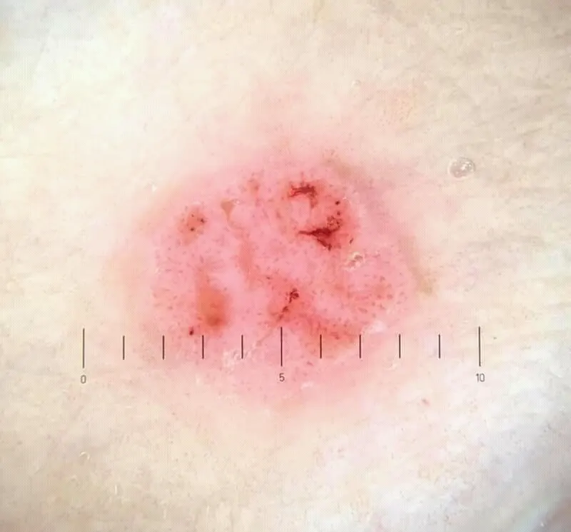
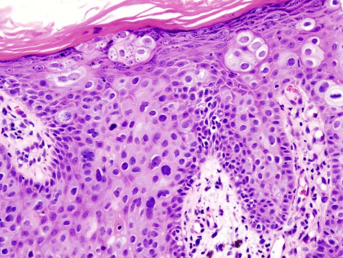
Actinic keratosis is more than a dry spot
It is a marker of chronic ultraviolet damage and often reflects a wider field of altered keratinocytes.
Bowen disease
This is squamous cell carcinoma in situ: abnormal cells remain within the epidermis. The key question is whether there is any invasive component.
Biopsy becomes especially important when: the lesion becomes firm or painful, ulcerates or bleeds, grows rapidly, is markedly hyperkeratotic, or does not respond to treatment.
Treatment
Actinic keratosis can be treated lesion by lesion or as a field. Bowen disease treatment depends on site, size, risk of invasion/recurrence and the need for reliable margin control.
European approach
Current guidance places actinic keratosis within the broader concept of UV-induced field cancerization.