Rare non-cutaneous melanoma

Mucosal melanoma

It arises on mucosal surfaces rather than the skin. The anatomic site determines symptoms, staging and local treatment, while its biology differs from typical cutaneous melanoma.

Nose and sinusesobstruction, recurrent bleeding, a mass
Oral cavitypigmented area, nodule, ulcer or bleeding
Anorectal regionbleeding, pain or a sense of a mass
Vulva / vaginaa lesion, pigment change or bleeding/discharge
Choose the site

Four different clinical pathways

Symptoms, local staging and surgical strategy depend on the anatomic site.

01

Nose and sinuses

Endoscopy, CT/MRI, REFCOR 2026, surgery and radiotherapy.

Open pathway →
02

Oral cavity

Pigmented and amelanotic disease, biopsy, pathology and IHC.

Open pathway →
03

Anorectal region

Endoscopy, pelvic MRI, pathology and function-conscious surgery.

Open pathway →
04

Vulva and vagina

Symptoms, MRI, pathology and an ethical approach to clinical imagery.

Open pathway →
For patients

What might I notice myself?

You cannot diagnose mucosal melanoma yourself. Most of these symptoms have benign causes. What matters is a new, persistent, unilateral, enlarging or bleeding mucosal change that deserves examination.

Nose and sinuses

Unilateral obstruction or recurrent bleeding

Persistent obstruction on one side, recurrent epistaxis, discharge, pain or a sense of a mass. Early symptoms are often nonspecific.

Examples and pathway →
Oral cavity

A macule, nodule or ulcer

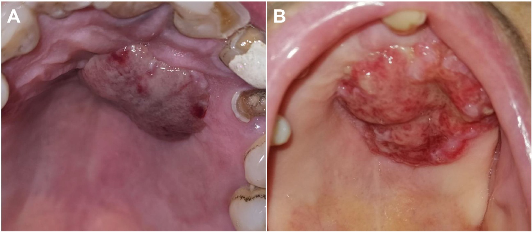
A new or enlarging dark area, irregular pigmentation, a nodule, ulcer or bleeding. Importantly, mucosal melanoma may be pink or red without obvious black pigment.

See real examples →
Anorectal region

Bleeding, pain or a sense of a mass

Blood in stool or on tissue, pain, a palpable lesion, tenesmus or bowel-habit change. These symptoms are far more commonly caused by other conditions, but persistent bleeding should not automatically be attributed to haemorrhoids.

Examples and pathway →
Vulva / vagina

A new lesion, itch or bleeding

Vulvar disease may cause a visible or palpable lesion, pigment change, itch, ulceration, pain or bleeding. Vaginal disease more often presents with bleeding/discharge, pain or a mass.

Signs and diagnosis →
When to arrange an examination: if a lesion is enlarging, does not heal, repeatedly bleeds, develops unexplained irregular pigmentation, or a symptom persists or progresses without a clear explanation. This does not mean cancer - it means the change deserves assessment.
If you remember only 3 things

This is not simply cutaneous melanoma in a different place

Mucosal melanoma is very rare. Persistent or unexplained symptoms are far more often caused by other conditions, but they deserve site-specific assessment.

1Not driven by sun exposure

Ultraviolet exposure is not the main explanation for its development.

2It may be non-pigmented

Amelanotic melanoma may be pink or red and may not resemble a typical dark melanoma.

3There is no single algorithm

Sinonasal, oral, anorectal and gynaecologic sites require different anatomic pathways.

Real clinical examples

Mucosal melanoma is not always black

Below are licensed clinical images from open-access publications, showing a spectrum from heavily pigmented to completely amelanotic lesions.
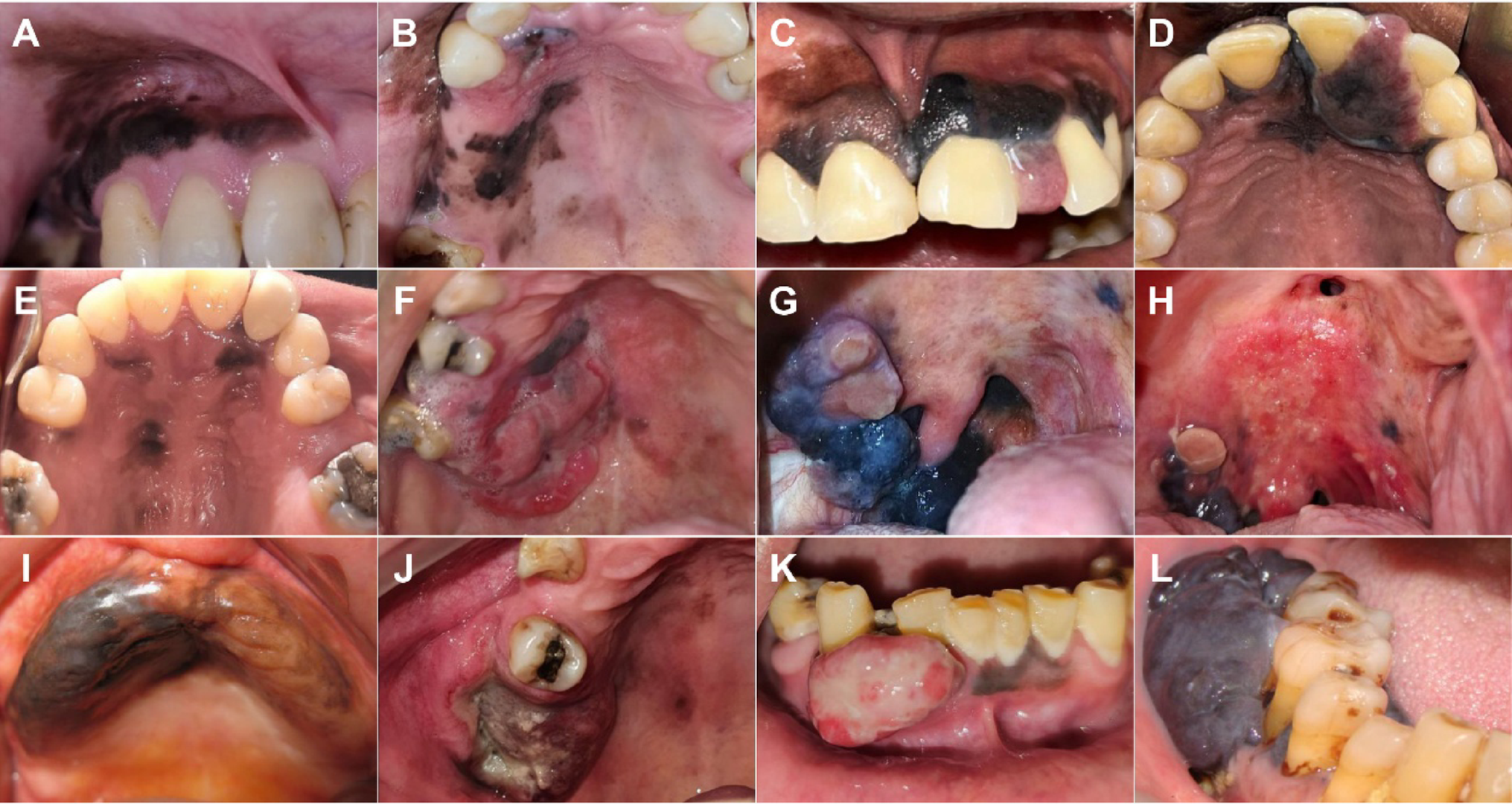
Clinical presentations of pigmented oral melanoma involving the palate, gingiva and other oral sites
Pigmented oral melanomaTwelve real clinical presentations, from macules to ulcerated exophytic tumours.de Arruda et al., Head Neck Pathol 2026 · source · CC BY 4.0
Two examples of amelanotic melanoma of the palate without visible dark pigmentation
Amelanotic melanoma of the palateTwo lesions without clinically visible melanin - an important reminder that colour does not exclude melanoma.de Arruda et al., 2026 · source · CC BY 4.0
Practical point: an atypical, enlarging, ulcerated or bleeding mucosal lesion may require biopsy whether black pigment is present or not.
Pathway

How the diagnosis is established

Two questions must be answered together: what is the pathology, and how far has the disease spread.

1Site-specific examination

ENT/endoscopy, oral-maxillofacial, colorectal or gynaecological examination depending on site.

2Biopsy

Histopathologic confirmation. Amelanotic or poorly differentiated tumours often require a broader immunohistochemical panel.

3Local imaging

MRI/CT is selected according to the anatomic site to define extent and relationships to critical structures.

4Systemic staging

Regional nodes and distant metastases are assessed; REFCOR 2026 recommends systemic staging at diagnosis for sinonasal mucosal melanoma.

5Molecular testing

In advanced disease, clinically relevant alterations including KIT, BRAF and NRAS are sought; broader NGS is useful when available.

Nose and paranasal sinuses

Endoscopy and CT

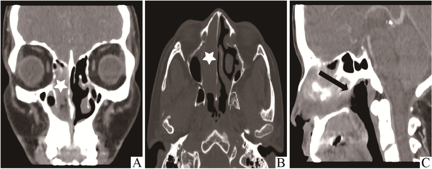
Sinonasal melanoma may present with nasal obstruction and recurrent epistaxis. Endoscopy demonstrates the lesion itself, while CT/MRI defines anatomic extent.
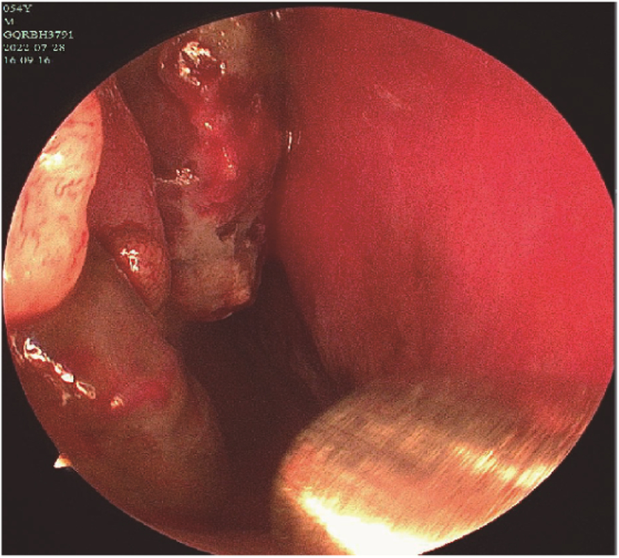
Endoscopic image of pigmented sinonasal mucosal melanoma in the nasal cavity
Endoscopic appearancePigmented tumour in the nasal cavity with evidence of prior bleeding.Tang et al., Diagnostic Pathology 2022 · source · CC BY 4.0
CT of sinonasal mucosal melanoma in coronal axial and sagittal planes
CT: local extentCoronal, axial and sagittal views demonstrate the mass and adjacent sinonasal structures.Tang et al., 2022 · source · CC BY 4.0
Anorectal melanoma

Endoscopy and pelvic MRI

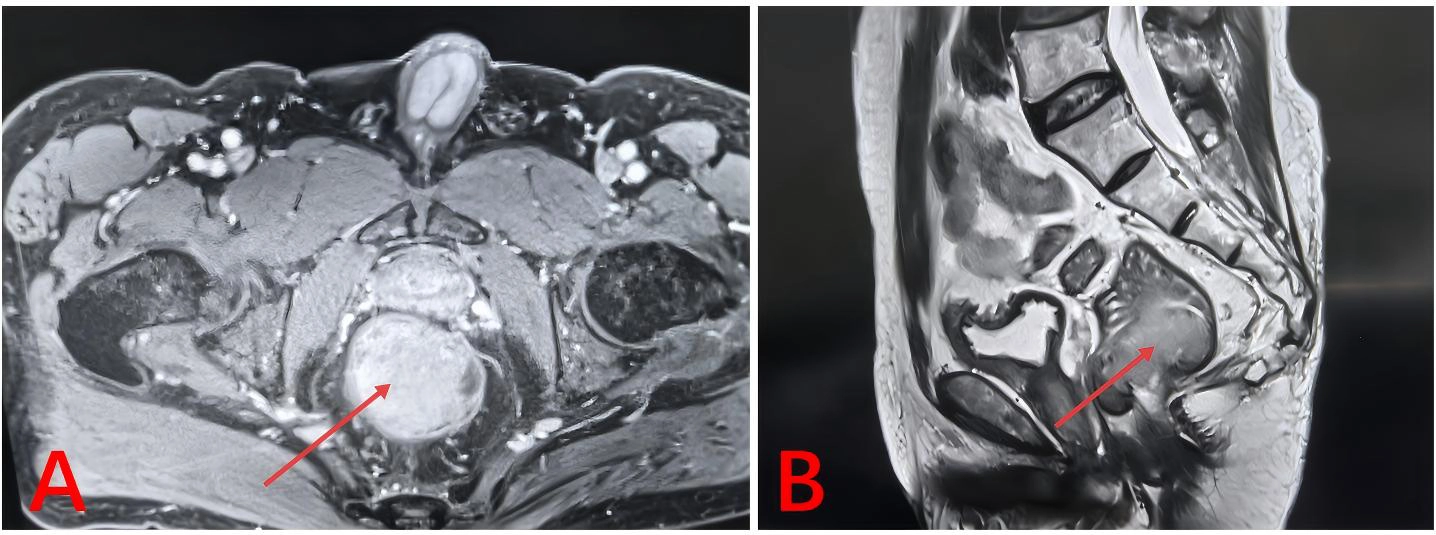
Bleeding or a sense of a mass may initially be attributed to benign anorectal disease. Endoscopy identifies the tumour, while MRI helps assess the sphincter and adjacent structures.
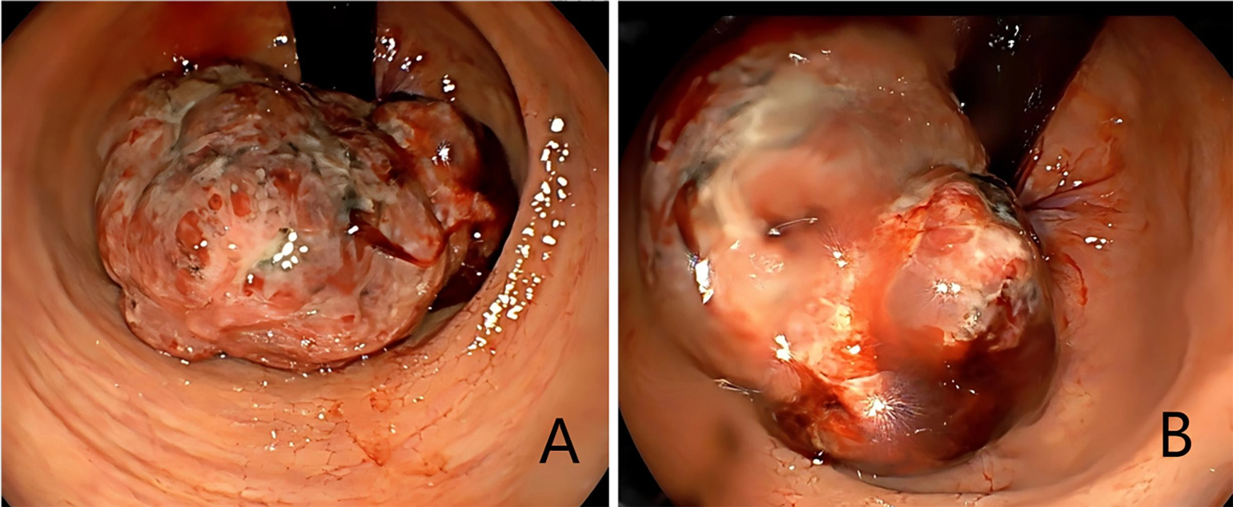
Colonoscopy showing a large irregular ulcerated anorectal melanoma
ColonoscopyLarge irregular ulcerated anorectal tumour.Wang et al., Front Oncol 2026 · source · CC BY
Axial and sagittal pelvic MRI of anorectal melanoma
Pelvic MRIAxial and sagittal imaging used to assess tumour size and relationships to surrounding structures.Wang et al., 2026 · source · CC BY
Vulva and vagina

For the patient layer - imaging rather than intimate clinical photographs

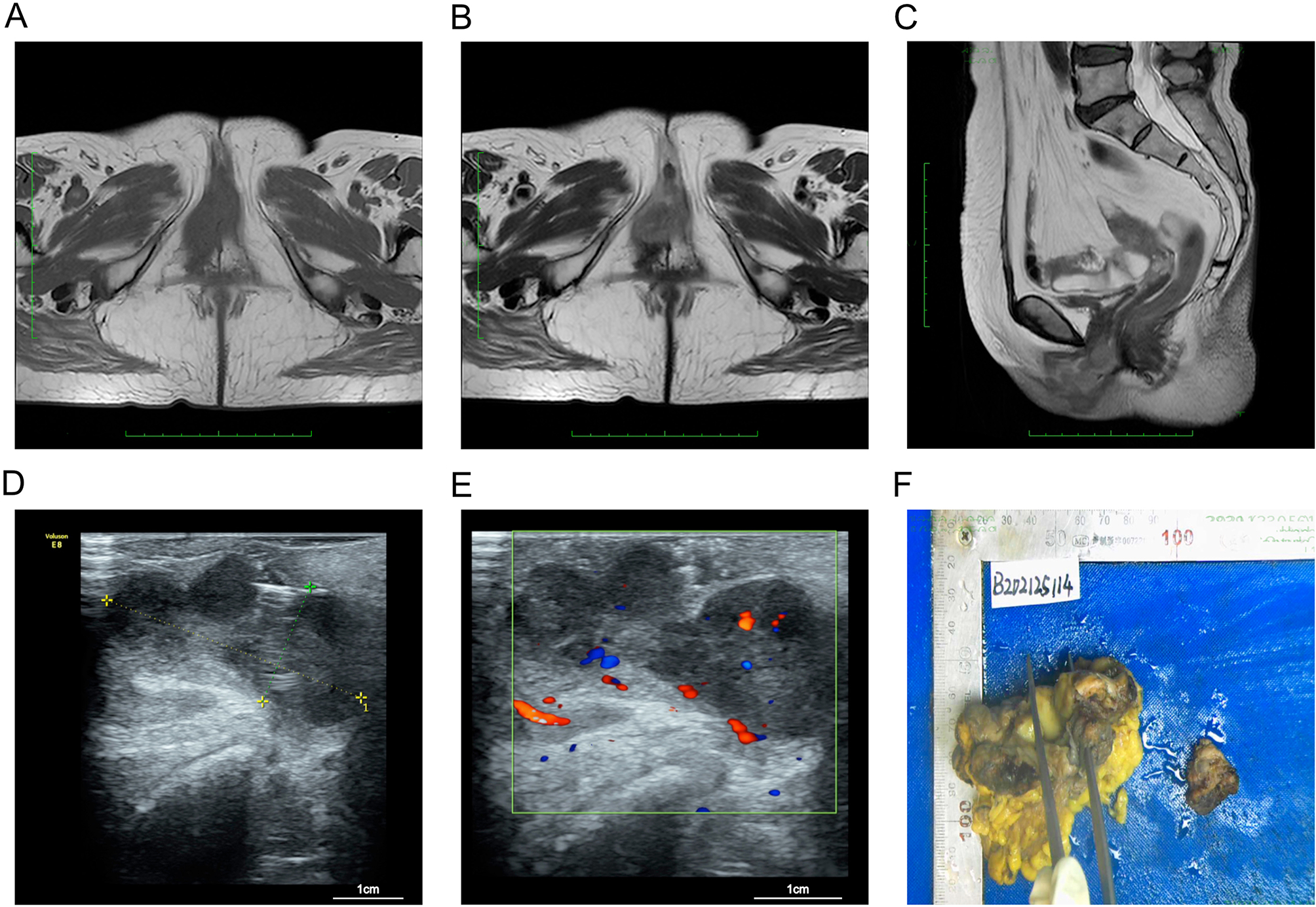
Even when reuse is legally permitted, we deliberately limit intimate clinical photography on the public patient layer. MRI can convey the diagnostic information without unnecessary exposure.
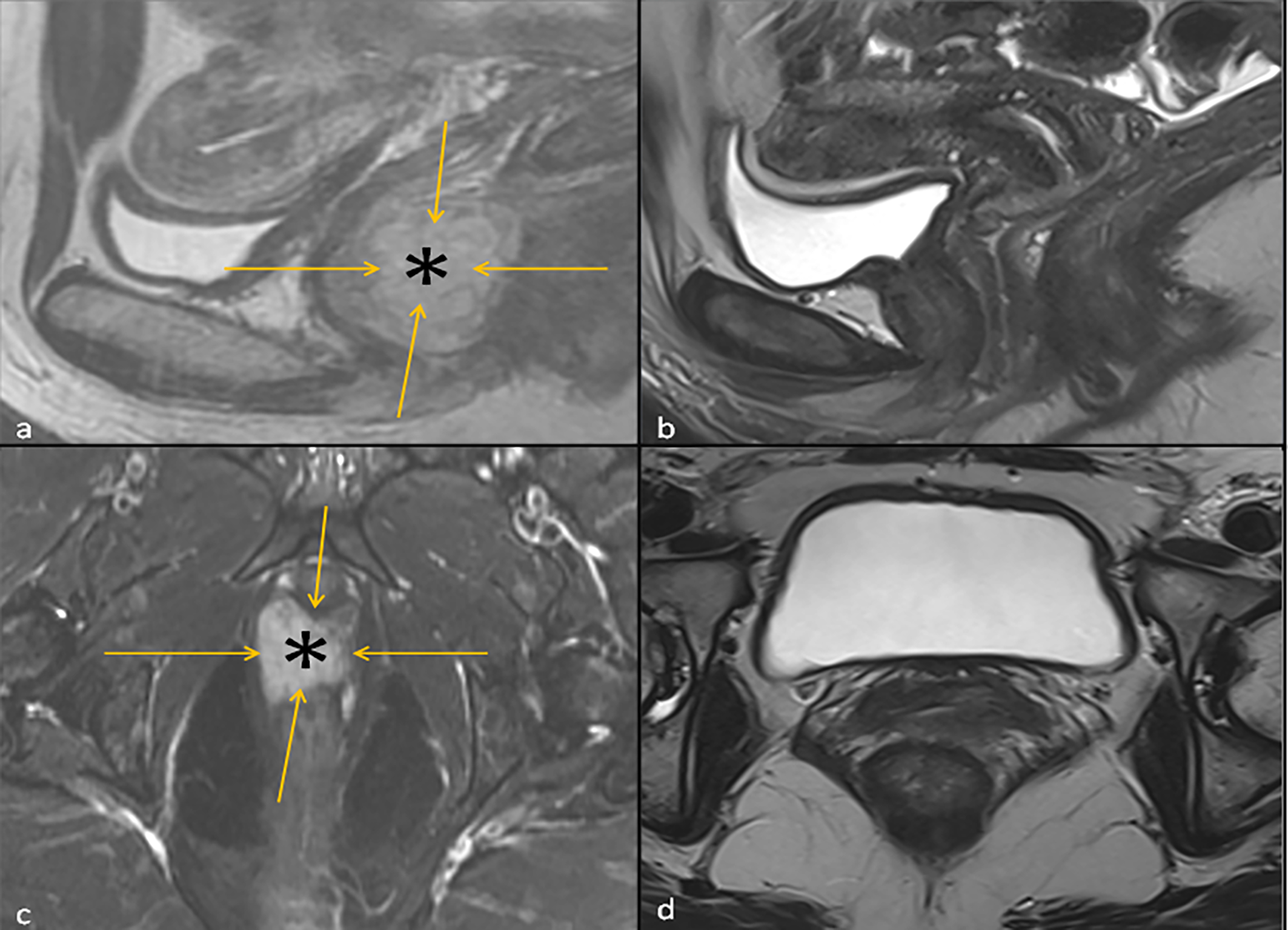
MRI of vaginal mucosal melanoma before and after immunotherapy
Vaginal melanoma: MRI before and after treatmentCase-report example showing the baseline tumour and subsequent radiologic response after ipilimumab + nivolumab. This illustrates an individual case and is not evidence for a universal strategy.Tarhini et al., Front Oncol 2022 · source · CC BY
MRI ultrasound Doppler imaging and surgical specimen in vulvar melanoma
Vulvar melanoma: MRI, ultrasound and specimenA multimodal imaging example; intimate clinical photography is intentionally not used on the patient page.Wang et al., BMC Cancer 2024 · source · CC BY 4.0
Treatment

Local control matters, but the decision should not be purely surgical

Because critical structures are often nearby, radical surgery may have a major functional cost. Management should incorporate anatomy, stage, tumour biology and systemic risk.

Localized diseaseSurgery remains the mainstay when adequate resection is feasible. The extent depends on the specific anatomic site.
RadiotherapyMay improve local control in selected scenarios, particularly in head-and-neck mucosal melanoma; its role is not identical to cutaneous melanoma pathways.
ImmunotherapyAnti-PD-1 and ICI combinations are used in advanced disease, but average response rates are lower than in cutaneous melanoma.
Targeted therapySelected KIT mutations and rare BRAF V600 alterations can change systemic treatment; KIT amplification alone is not equivalent to an activating mutation.
Clinical trialsParticularly important because the subtype is rare and dedicated randomized evidence is limited.
Professional layer

Full atlas + staging + pathology + molecular + systemic evidence

A separate clinician hub covers pathology and IHC, site-specific staging, nodal management and SLNB, surgery, radiotherapy, KIT/BRAF/NRAS/NF1, direct mucosal cohorts and investigational strategies in 2025–2026.

Open clinician hub
Media policy: only material with a clear reuse licence is displayed (primarily CC BY 4.0 / CC BY). Every image links to the original publication with attribution. NC/ND or uncertain-rights material is intentionally excluded.