Oral mucosal melanoma
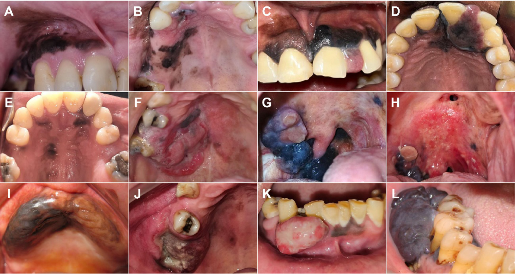
It may appear as a dark macule, nodule or ulcer, but some tumours contain little or no clinically visible pigment. Persistent atypical mucosal lesions should therefore not be judged by colour alone.
Not only a dark spot
Self-checking does not replace clinical examination, but oral mucosal changes may be noticed during tooth brushing or a dental visit.
Brown, black, bluish or irregular pigmentation, particularly if it enlarges.
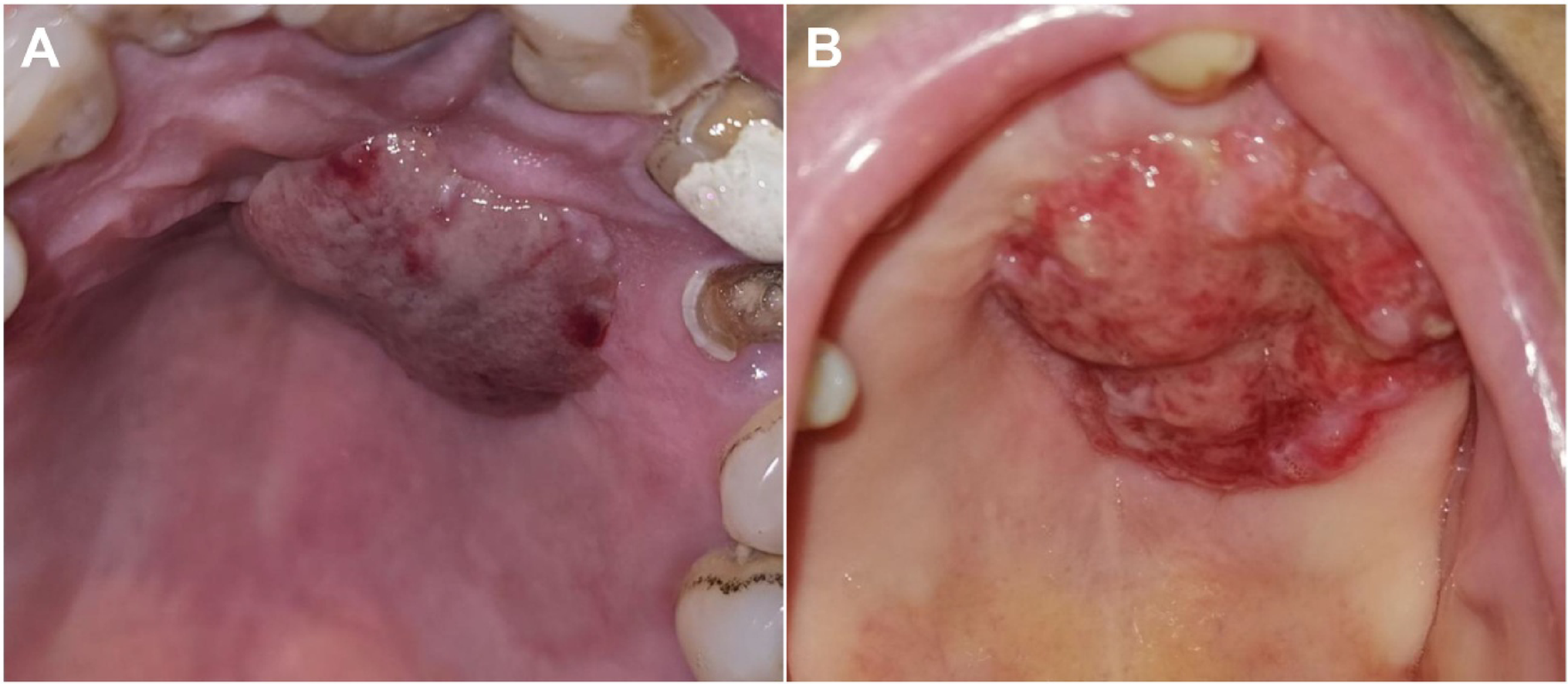
Amelanotic melanoma may have little or no visible pigment.
A new raised lesion on the palate, gingiva or another oral mucosal site.
A lesion that does not heal, is repeatedly traumatized or bleeds deserves assessment.
A strong open clinical image series
The de Arruda et al. series shows a true spectrum rather than a single stereotypical presentation.
Examination → biopsy → pathology → staging
The pathway must answer both what the lesion is and how far disease extends locally and systemically.
Oral/maxillofacial or oncologic assessment documenting site and extent.
Adequate tissue for morphology and IHC; amelanotic tumours can mimic other malignancies.
Selected according to anatomy to assess bone, soft tissues and regional nodes.
Local control must be balanced against speech, swallowing, mastication and reconstructive options.
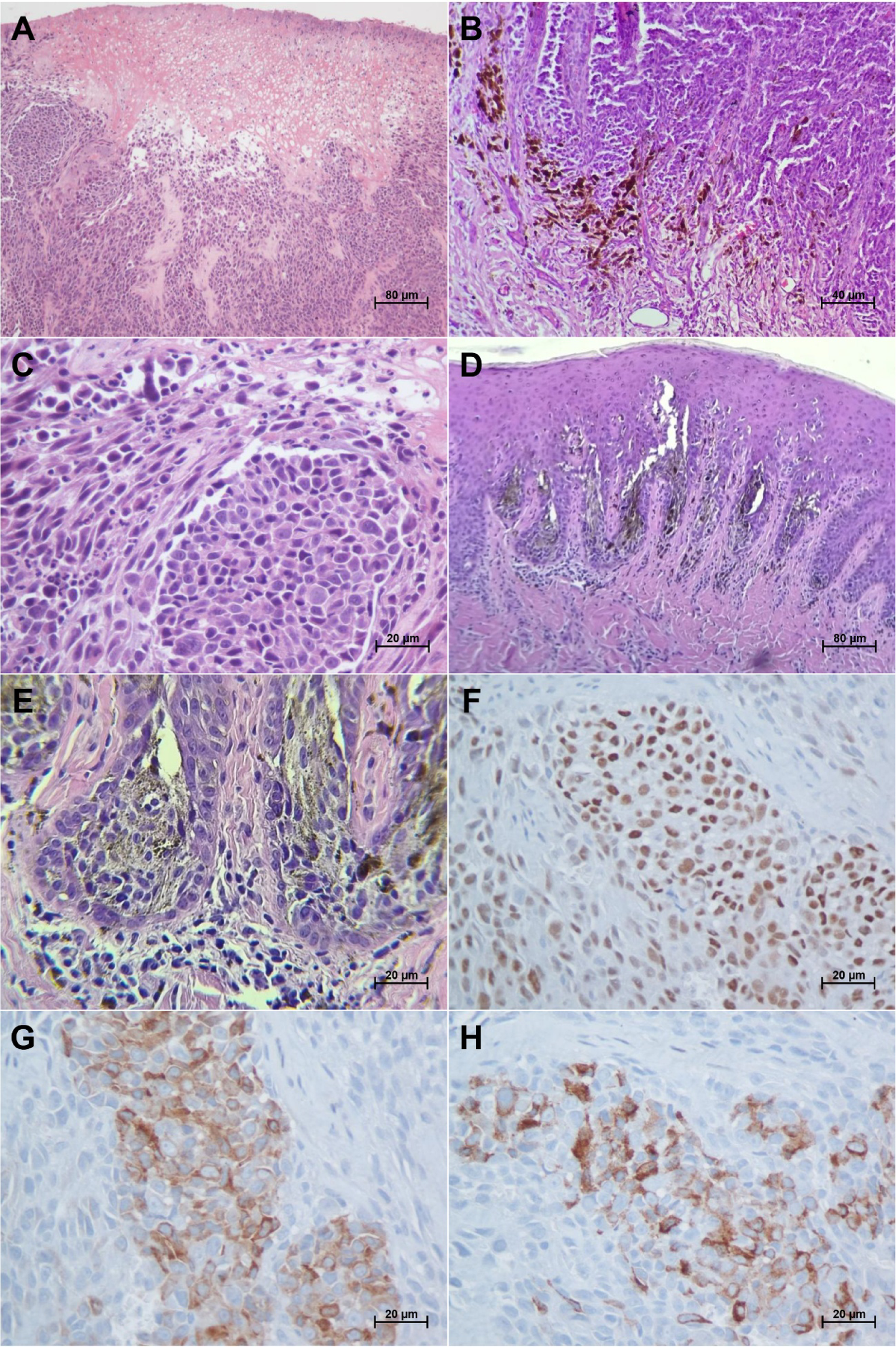
When pigment is limited, immunohistochemistry is particularly important
SOX10, S100, Melan-A and HMB45 are used as part of a panel in morphologic context; no single marker should be interpreted in isolation.