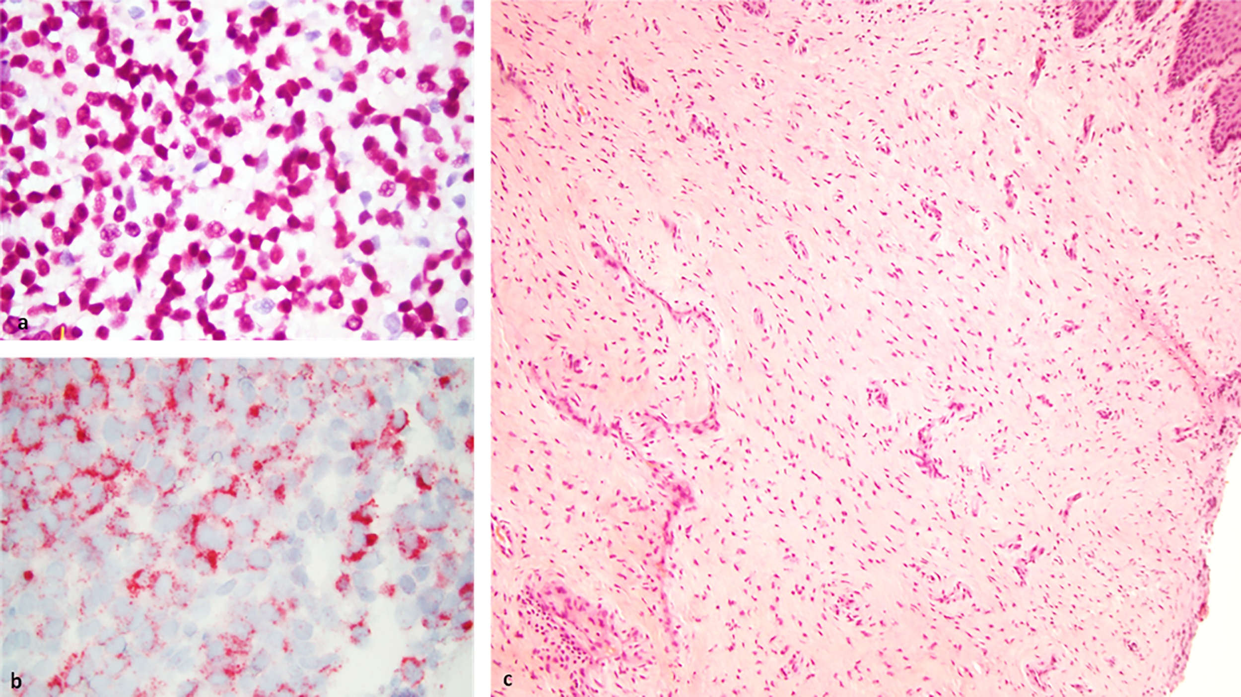
Vulvar and vaginal melanoma
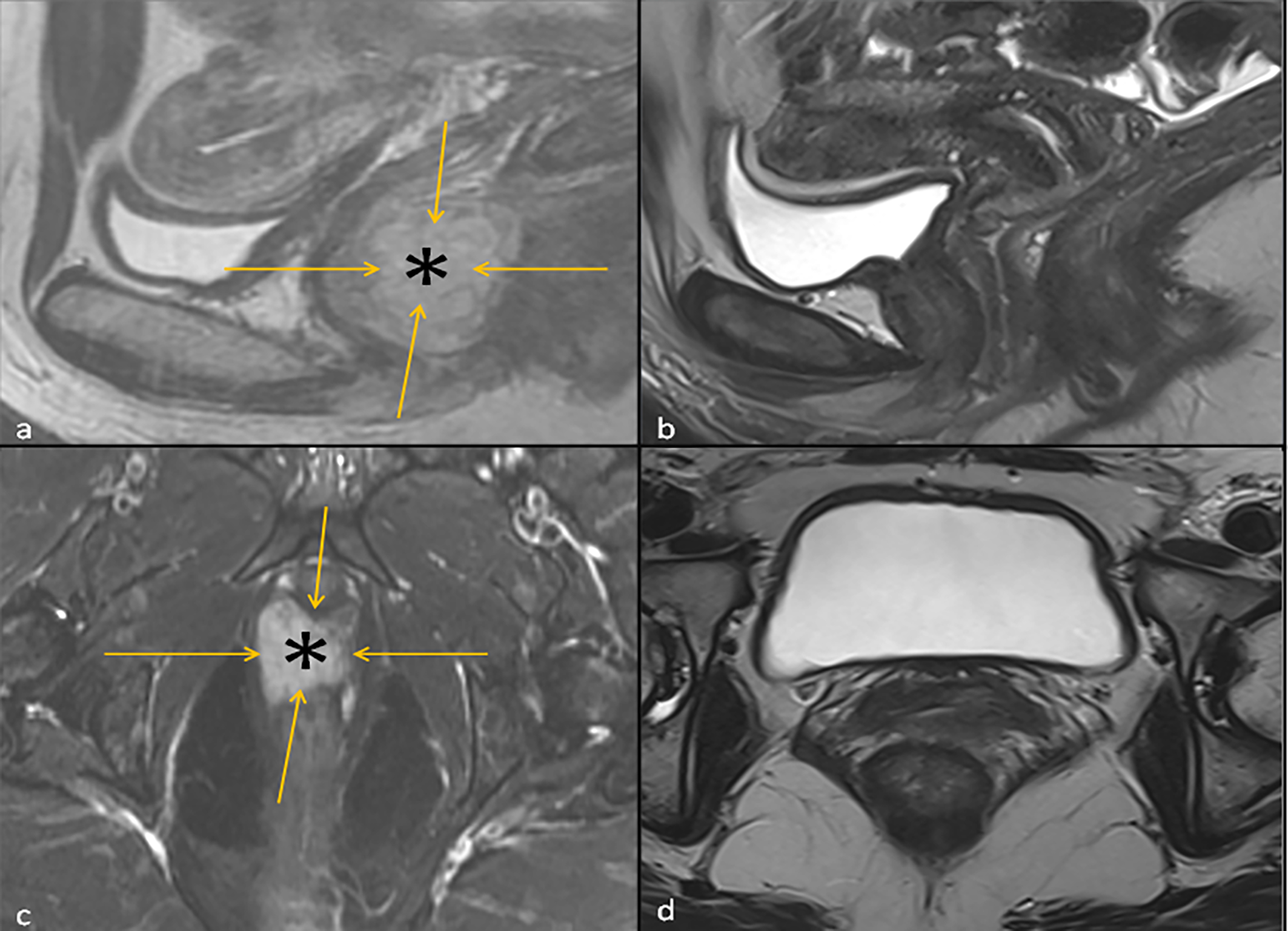
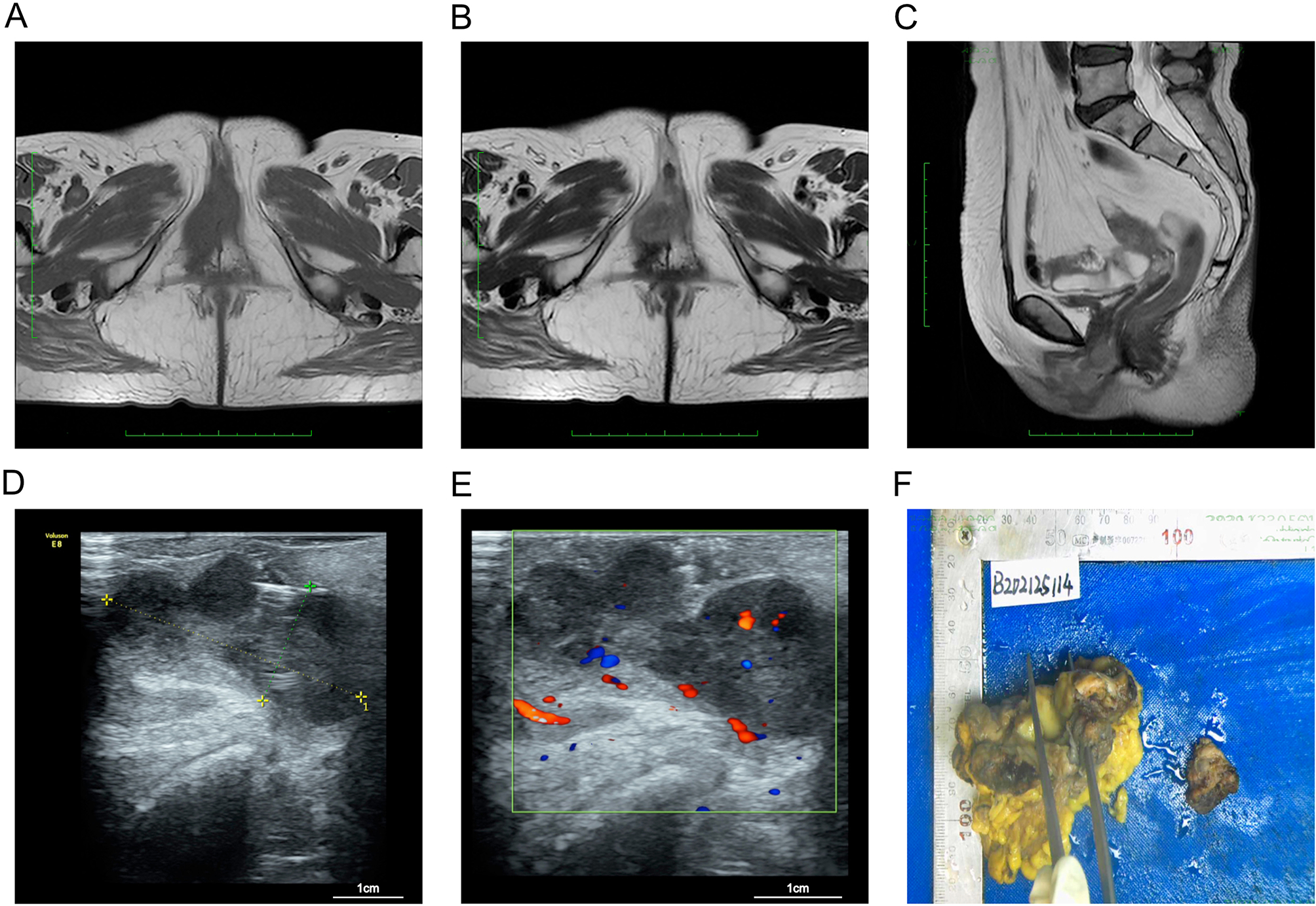
Rare malignancies with different anatomy but the same need for accurate pathology, local staging and assessment of systemic risk. On the public patient layer we deliberately favour MRI and pathology rather than intimate clinical photographs.
Symptoms depend on the site
Most such symptoms have benign causes, but persistent change deserves gynaecologic assessment.
Pigmented or without obvious pigmentation.
A new or evolving asymmetric area.
Especially without a clear benign explanation.
Nonspecific symptoms interpreted together with examination.
MRI helps plan local treatment
The patient page can communicate diagnostic information without unnecessary exposure of intimate clinical photography.
Amelanotic disease can be diagnostically challenging
Morphology is interpreted together with immunohistochemistry and clinical context. The clinician section separately addresses SOX10/S100/Melan-A/HMB45, PRAME and differential diagnosis.