Selected superficial defects
Direct closure, secondary intention or full-thickness skin graft when contour and nearby structures will not be distorted.
Nasal reconstruction is three-dimensional. Skin cover alone may be insufficient when cartilage support or internal lining is lost; airway function and contour must be considered alongside cancer control.
Tip, dorsum, sidewall, ala, columella, and adjacent cheek or medial canthus.
Skin and soft tissue only, or full-thickness loss involving cartilage or mucosal lining.
Is the alar rim, external nasal valve and structural framework preserved?
Confirm the oncologically appropriate defect and consider margin control before definitive closure.
Direct closure, secondary intention or full-thickness skin graft when contour and nearby structures will not be distorted.
V-Y advancement, rotation, transposition or dorsal nasal flaps depending on laxity, vascularity and eyelid/canthal traction.
Options may include melolabial, nasolabial or paramedian forehead flaps, auricular cartilage support and restoration of lining.
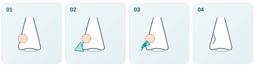
Record the subunit, oncological margins and three-dimensional tissue loss.
Assess donor-tissue laxity, tension vectors and whether cartilage support is needed.
Advance a flap or rebuild lining, framework and cover as indicated.
Assess symmetry, nostril patency and later functional outcomes.
Each panel represents a separate patient; images from different cases are not presented as one sequence. Immediate repair is not labelled as a late outcome.
A nasal defect after oncological excision was reconstructed using a pedicled melolabial (nasolabial) flap recruited from the adjacent cheek. The surgeon provided three sequential clinical photographs from the same case.
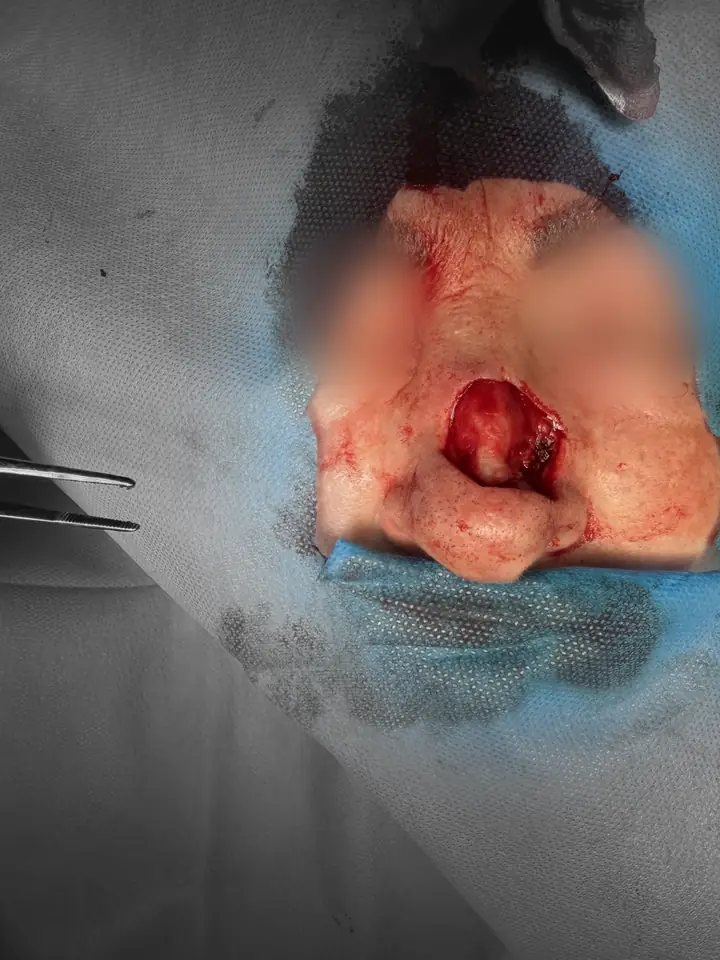
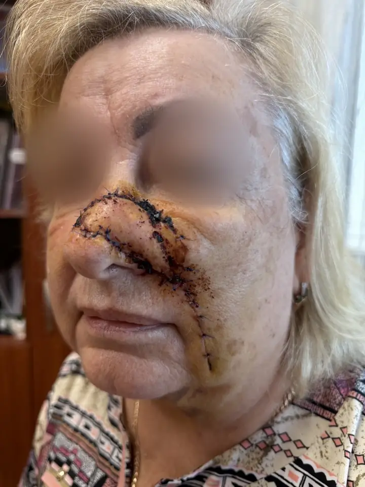
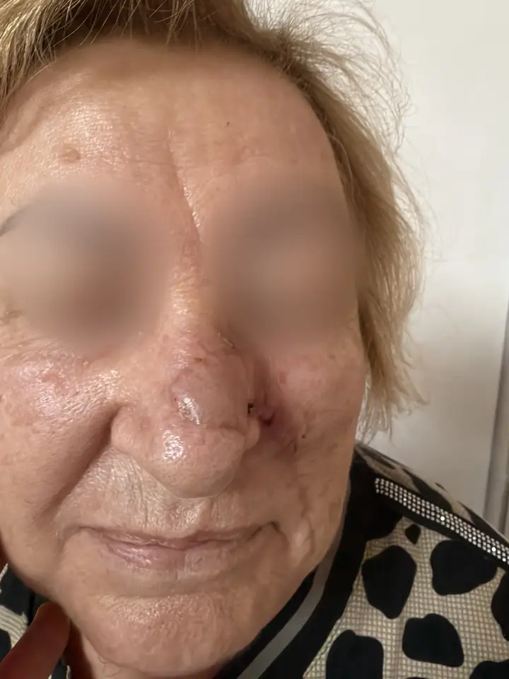
Why a staged flap: the melolabial region provides nearby tissue with similar colour and thickness while maintaining perfusion during flap integration. The precise tumour histology, defect measurements, resection margin status and requirements for cartilage support or nasal lining are not established by this image set and are not inferred.
Real clinical case from the surgeon's archive. Publication authorised by the author; the eye regions were softly blurred, and the initial image's background was desaturated. The surgical defect, flap and postoperative changes have not been altered.
The defect was reconstructed with a bilobed local flap following micrographic margin control. The existing source article explicitly documents one Mohs stage and one-year follow-up. No pre-excision tumour photograph is available in this case set.
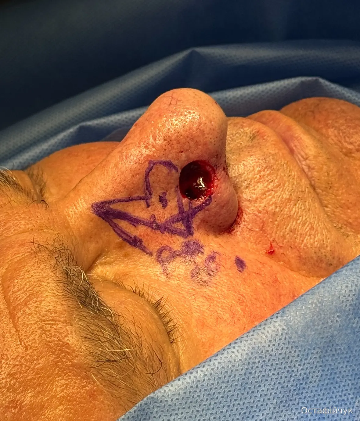
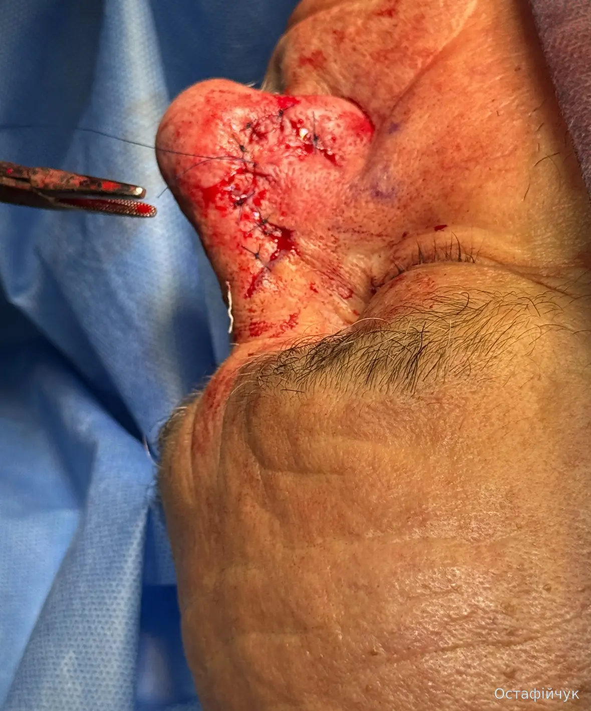
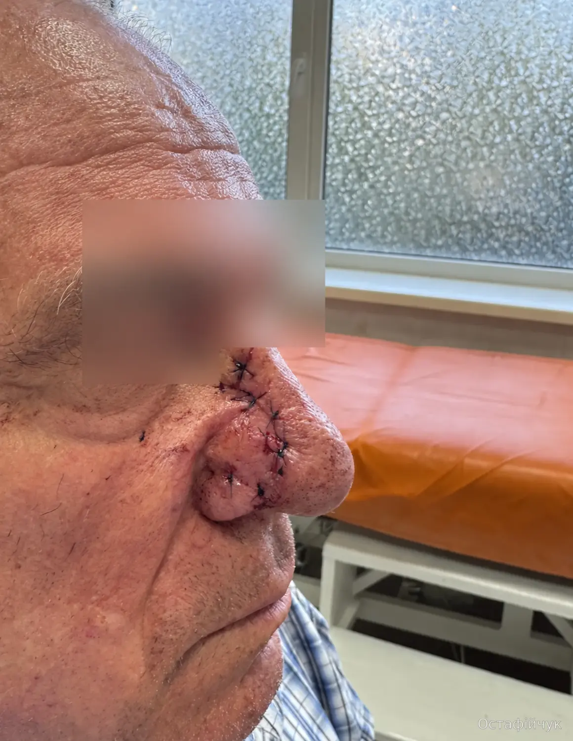
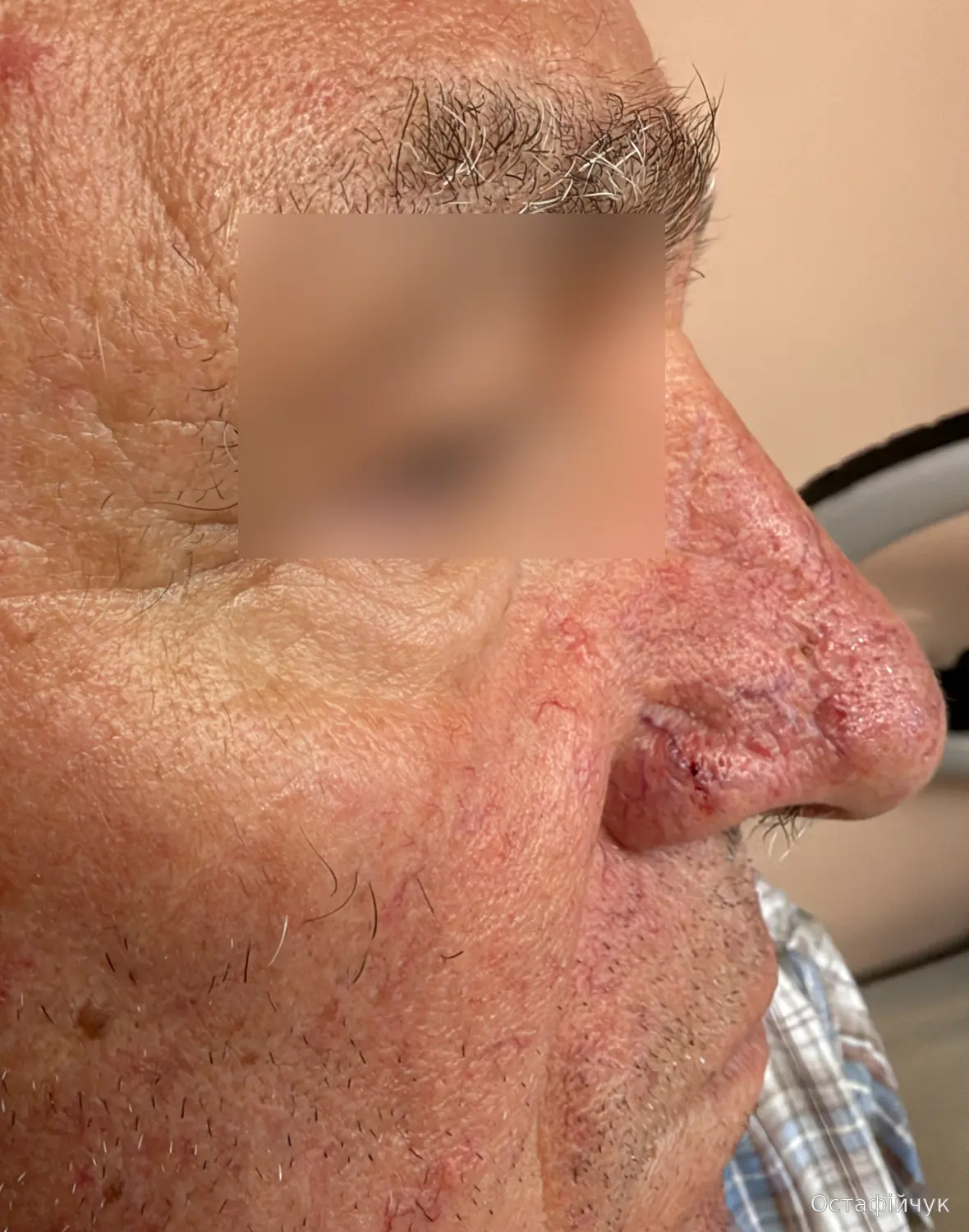
Clinical rationale. A bilobed flap recruits adjacent tissue with similar qualities. Alar traction and cartilage support remain relevant considerations; the flap choice cannot be inferred from diameter alone.
Source: the author's already published clinical archive; images reproduced from existing site resources. This is a real clinical sequence, not an illustrative simulation.
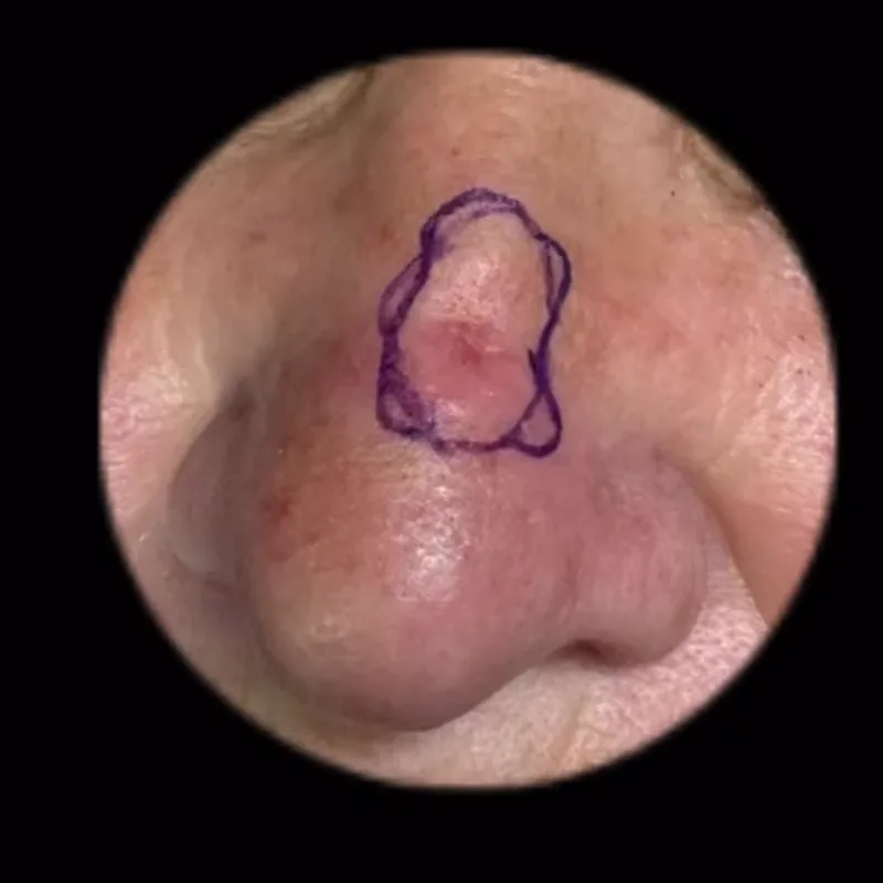
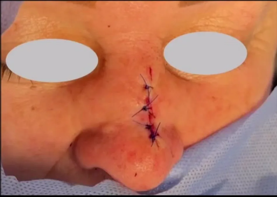
Separate material from the author's reconstruction resource. The images show marking and immediate closure, without a documented long-term scar result or full margin-assessment sequence in this set.
Source article and original captions →
Source: the author's already published clinical archive; images reproduced from existing site resources. This is a real clinical sequence, not an illustrative simulation.
Previously published, permission-cleared author material contains nasal marking and immediate local reconstruction. These examples illustrate geometry rather than a universal surgical plan.
Existing author reconstruction atlas →Published reviews describing anatomical principles and available options. Technique selection remains patient- and surgeon-specific.