Selected small helical defects
Direct wedge closure when tension and cosmetic distortion are acceptable.
A small loss of auricular cartilage can cause disproportionate contour changes. Reconstruction depends on the helical rim, antihelix, concha and any involvement of the external auditory canal.
Is the chondrocutaneous margin disrupted and is advancement possible without unacceptable ear reduction?
Assess concave surface, perichondrium and the graft or flap recipient bed.
Consider how to preserve height, structural support and symmetry.
Potential EAC narrowing or tumour extension requires separate specialist planning.
Direct wedge closure when tension and cosmetic distortion are acceptable.
Antia–Buch chondrocutaneous advancement can restore a selected rim defect, within anatomical size limits.
A revolving-door or other postauricular flap, graft, staged repair or cartilage reconstruction may be considered based on depth and location.
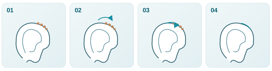
Map the rim, cartilage and auditory canal before closure.
Plan a method that preserves the natural ear contour and support.
Mobilise a local or postauricular flap with appropriate vascularity.
Assess symmetry, auricular position and canal patency.
These photographs come from the skin cancer archive and depict two different patients before treatment. They are not a before-and-after pair or a documented Antia–Buch reconstruction. A reconstruction cannot be inferred retrospectively from the tumour photo alone.
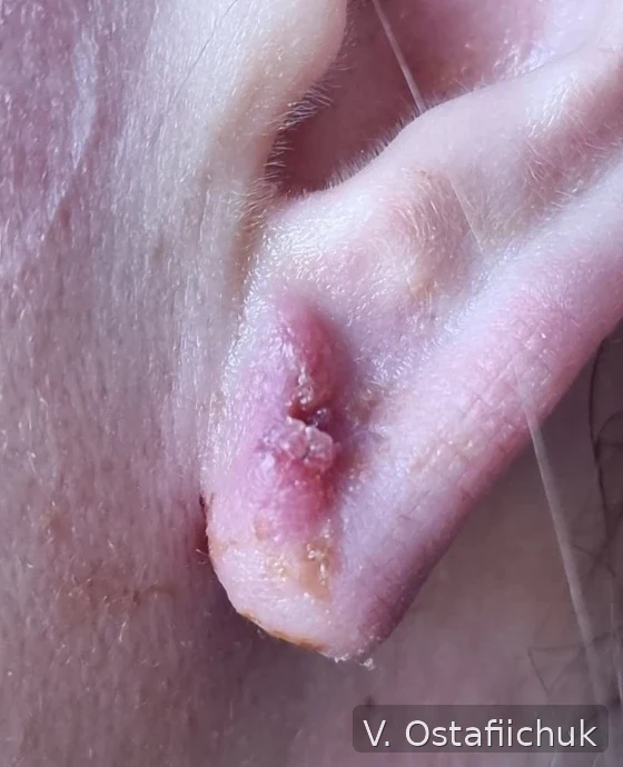
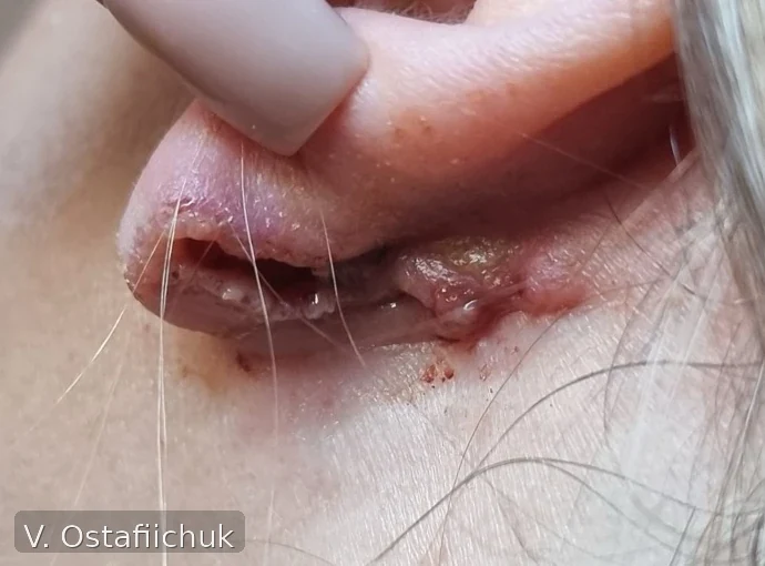
Source: skin cancer knowledge centre →
Source: the author's already published clinical archive; images reproduced from existing site resources. This is a real clinical sequence, not an illustrative simulation.
The existing skin cancer centre includes educational images of auricular tumours, but these are not a documented Antia–Buch reconstruction series. No patient outcomes are invented.
Existing author reconstruction atlas →Published reviews describing anatomical principles and available options. Technique selection remains patient- and surgeon-specific.