Small defects
Consider tension-free direct closure or selected local cover for superficial defects.
Eyelid reconstruction must preserve more than appearance: corneal protection, lid-margin position, horizontal support and lacrimal drainage are decisive outcomes.
Anterior: skin and orbicularis; posterior: tarsus and conjunctiva.
Estimate the proportion of eyelid margin lost and available tissue laxity.
Assess medial and lateral canthal support and the lacrimal system.
Plan lid-globe apposition and minimise ectropion or exposure keratopathy.
Consider tension-free direct closure or selected local cover for superficial defects.
Tenzel semicircular flap or other combined techniques when anatomically appropriate; support and tension vectors matter.
Hughes tarsoconjunctival flap for posterior lamella with anterior coverage as needed; a Mustardé cheek rotation flap may be an option for anterior cover in selected cases.
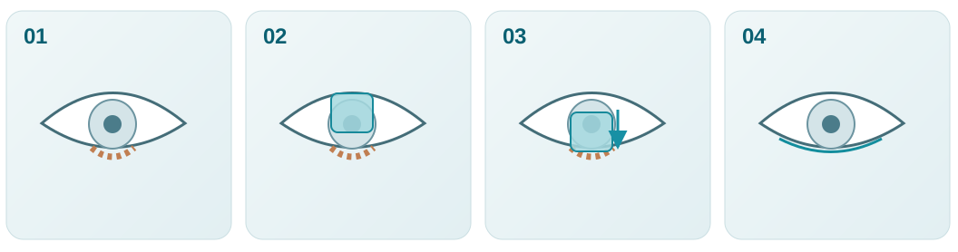
Identify anterior and posterior lamellar loss and eyelid-margin involvement.
Address posterior support, anterior cover and canthal stability separately.
Rebuild each lamella as appropriate; Hughes is a staged procedure.
Evaluate eyelid position, corneal protection and healing over time.
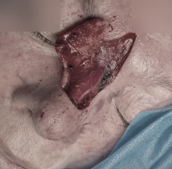
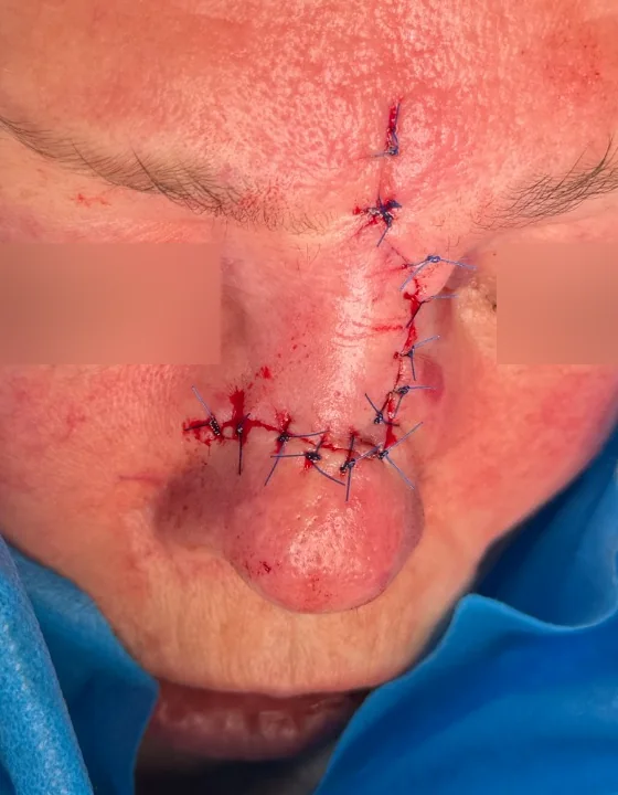
Periocular reconstruction is not necessarily a reconstruction of the eyelid margin. We describe what each source actually shows and do not mislabel these photographs as a Hughes procedure.
These two photographs from an existing published case show the post-excision defect and immediate local flap reconstruction. The source does not establish the precise technique or a long-term functional outcome.
Original article and captions →
Source: the author's already published clinical archive; images reproduced from existing site resources. This is a real clinical sequence, not an illustrative simulation.
The previously displayed single-image periocular case has been removed. For an accurately documented staged nasal reconstruction, see the two-stage melolabial flap clinical series.
Previously published author material includes periocular reconstructions. The clinical examples above now accompany the conceptual teaching schemes.
Existing author reconstruction atlas →Published reviews describing anatomical principles and available options. Technique selection remains patient- and surgeon-specific.