Uveal melanoma: symptoms and how it is detected
Uveal melanoma may cause no symptoms for a long time. It helps to know which changes deserve an eye examination and how an ophthalmologist confirms or rules out a suspicious lesion.

What a person may notice
Multimodal assessment - not one test
Visual acuity, slit-lamp examination, dilated pupil and fundus examination.
Documents size, colour, location and change over time.
B-scan assesses shape, thickness and internal reflectivity; ESMO recommends high-frequency UBM for ciliary-body lesions.
Can show subretinal fluid, orange pigment and other features when clinically relevant.
Used to evaluate local extent and, after diagnosis, to investigate metastatic disease when indicated.
A typical lesion is often diagnosed clinically. Biopsy is discussed for diagnostic uncertainty and/or prognostic molecular profiling.
How iris melanoma can look
Not every melanoma is dark and not every lesion has the same morphology.
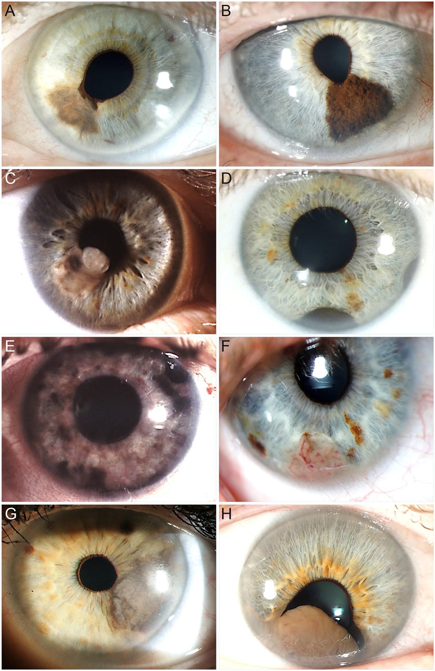
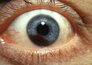

A photograph cannot rule disease in or out
Choroidal and ciliary-body melanoma are usually not visible externally. Iris melanoma can also be weakly pigmented or amelanotic.
MRI series from one real choroidal melanoma case
The same lesion across planes and sequences.
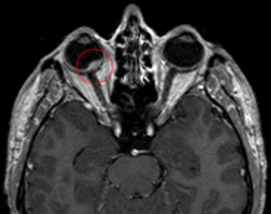
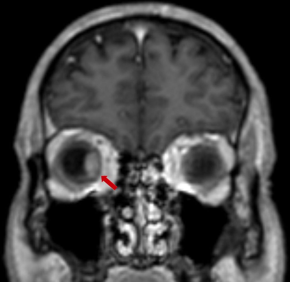
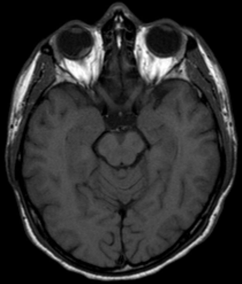
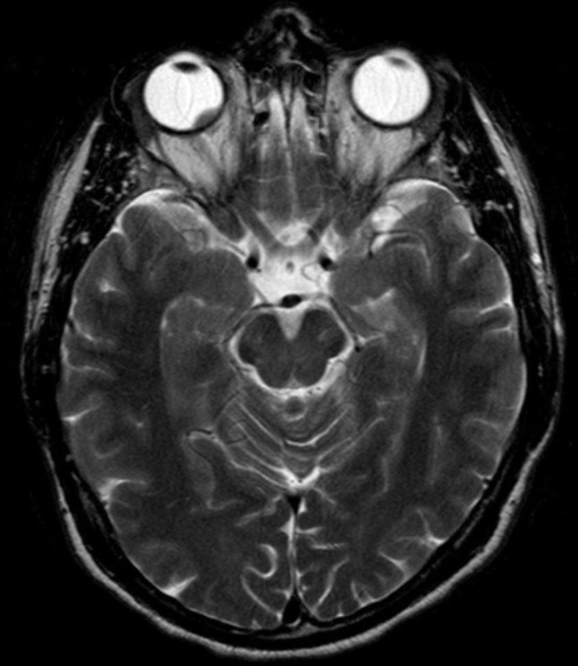
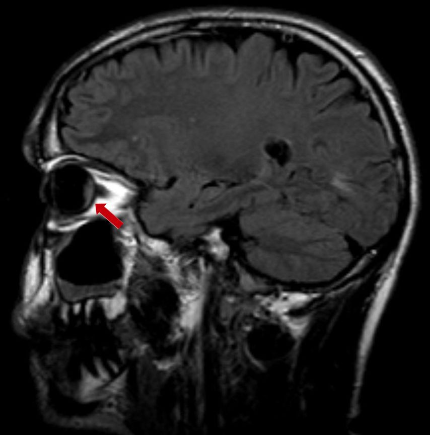
Author: Hellerhoff. Wikimedia Commons. CC BY-SA 4.0. Images are displayed without clinical alteration.
For clinicians: TFSOM-DIM features associated with nevus transformation
This is a risk framework, not an automatic diagnosis.
For clinicians: open-access multimodal clinical sets
For clinicians: pathology / gross specimen
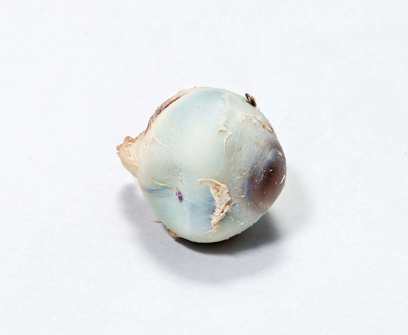

Research video: UM cell transendothelial migration
Basic-science video, not patient diagnostic imaging.
Onken M, Li J, Cooper J. PLOS ONE 2014. CC BY 4.0.
- ESMO–EURACAN Clinical Practice Guideline. ESMO Open. 2026.
- NCI PDQ — Intraocular (Uveal) Melanoma, Patient Version.
- American Cancer Society — Signs and Symptoms of Eye Cancer.
- American Cancer Society — Tests for Eye Cancer.
- Shields CL, et al. Multimodal imaging features predictive of choroidal nevus transformation. 2019.