Surveillance after uveal melanoma
Follow-up should reflect risk. Imaging interval and modality depend on tumour biology, clinical context and local availability.

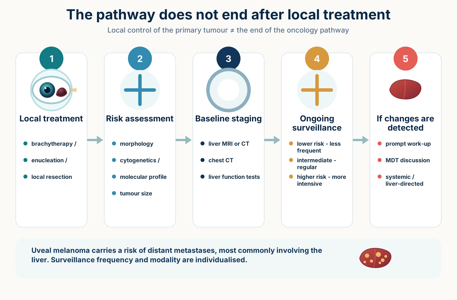
What ESMO–EURACAN recommends in 2026
After local treatment, follow-up has two parallel tracks: monitoring the treated eye and risk-adapted surveillance for systemic progression.
Then annually. Colour photography is highly effective for local recurrence; ocular US is valuable when the tumour is not fully visible.
A suspicious finding should trigger liver MRI.
The plan should be coordinated through a reference centre using clinical and genetic risk factors.
What the 2026 North American Delphi consensus adds
For intermediate risk, expert consensus favours contrast-enhanced abdominal MRI over US and CT: every 3–6 months for 5 years, then annually to at least 10 years. For high risk: every 3–6 months in years 1–5 and every 6–12 months in years 6–10; chest CT is combined with hepatic imaging but may be less frequent.
For clinicians: practical surveillance points
- PET/CT is not recommended for routine surveillance regardless of risk class in the Delphi consensus.
- Routine laboratory surveillance is unlikely to add diagnostic sensitivity when high-quality imaging is performed.
- With hepatic steatosis or body habitus that limits ultrasound, contrast-enhanced CT or MRI is preferred.
- 15-GEP/PRAME, chromosome 3/8, BAP1 and clinical stage are prognostic; they do not automatically select a systemic drug.