Melanoma of the palm, sole or nail unit
Acral melanoma arises on palms, soles and the nail apparatus. It is often unrelated to ultraviolet exposure, can look atypical and may therefore be diagnosed later.
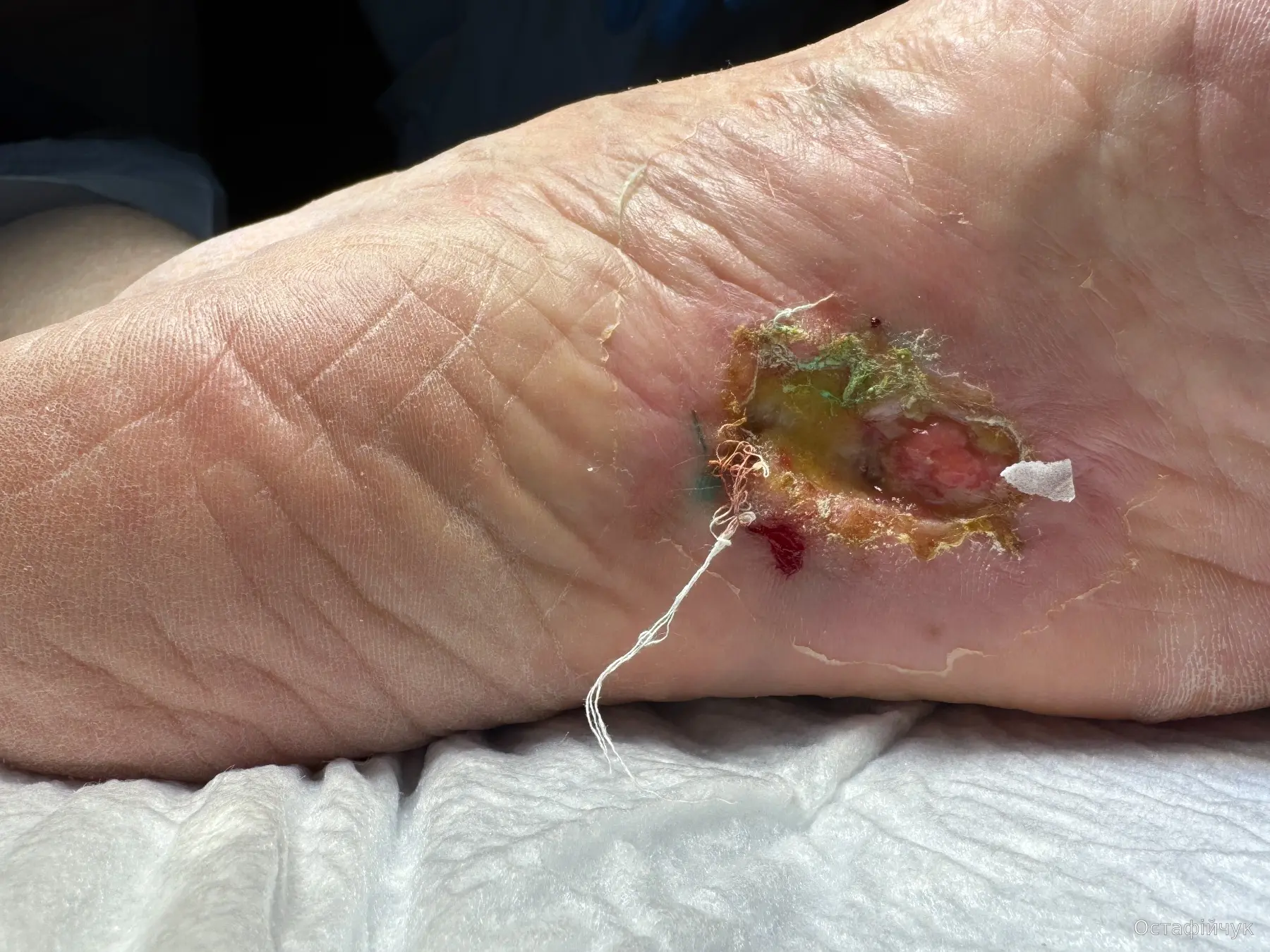
What should be examined
None of these signs means cancer automatically. They mean the lesion deserves an in-person assessment.
Especially if asymmetric, colour-variegated or gradually enlarging.
Persistent ulceration, bleeding or thickening on the foot should not be treated for months without tissue diagnosis.
Particularly when there is no clear injury, it is widening or extends onto periungual skin.
Acral melanoma can be weakly pigmented or amelanotic.
How diagnosis and treatment are planned
The goal is an adequate diagnosis without losing the opportunity for function-preserving definitive surgery.
Assess lesion architecture, nail unit involvement, ulceration and regional nodes.
Technique depends on size and site; the specimen must allow reliable assessment of invasion and Breslow thickness.
Generally follows cutaneous melanoma principles; SLNB is discussed according to thickness, ulceration and risk.
Achieve adequate oncologic control while preserving function whenever safely possible.
Skin grafts, local flaps or other approaches are tailored to weight-bearing and functional needs.
High-risk and advanced disease decisions depend on stage and molecular profile, with the caveat that acral melanoma is underrepresented in many pivotal trials.
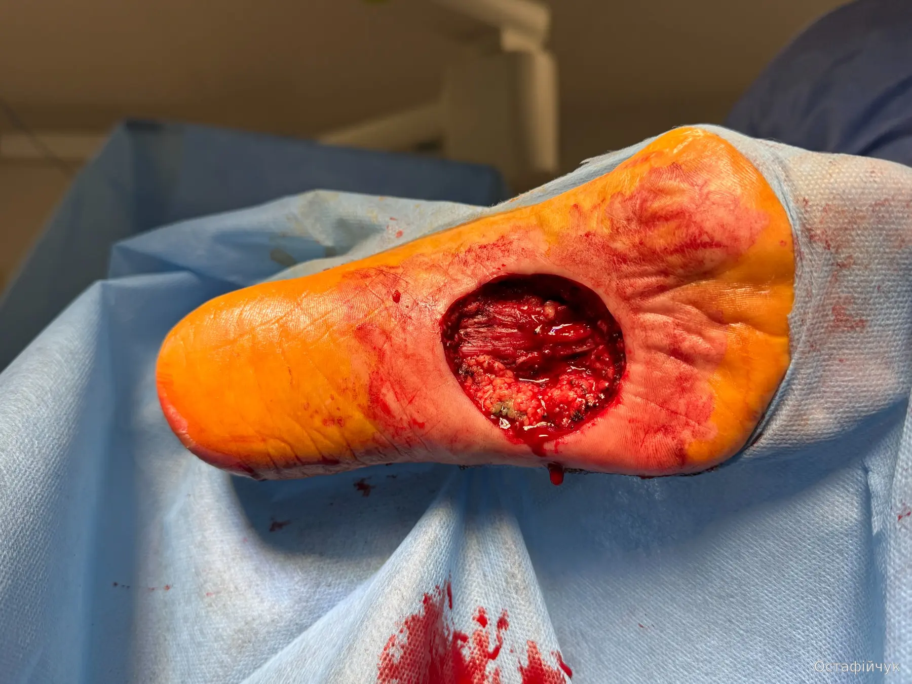
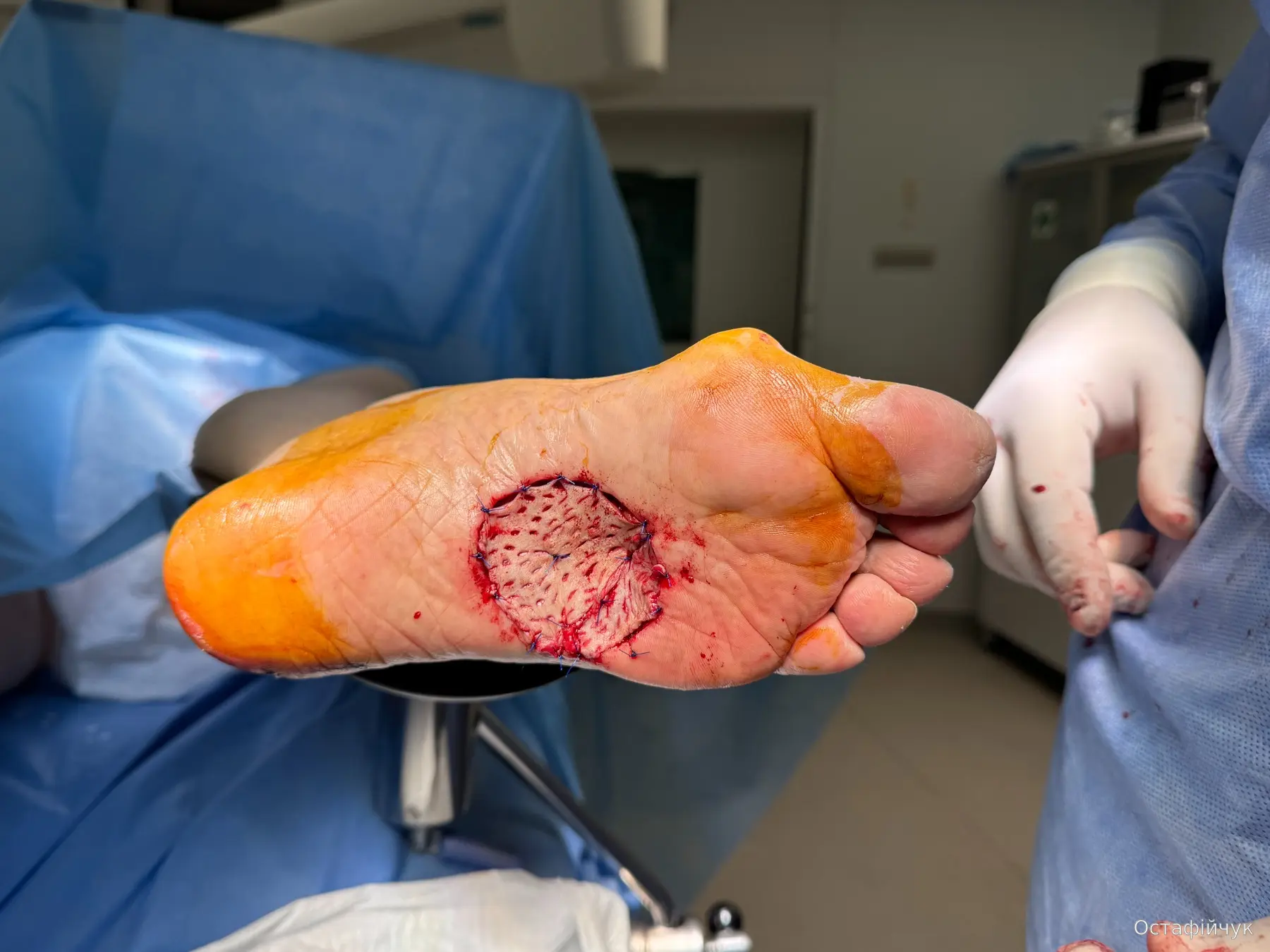
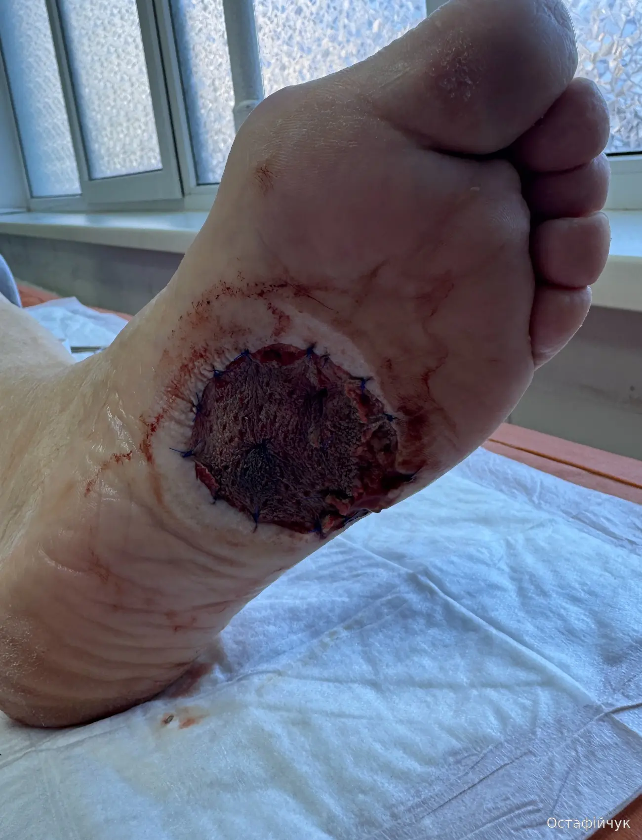
From a plantar tumour to reconstruction
Wide excision, free skin graft and negative-pressure wound therapy for 7 days.
What is distinct about acral melanoma
Lower mutational burden, more structural/copy-number change, a different KIT/NF1/TERT spectrum and lower representation in large Western melanoma trials. Surgical principles are largely extrapolated from cutaneous melanoma, but the anatomy of the sole, palm and nail unit requires specific functional planning.