Melanoma SLNB quality checklist
Seven checkpoints from indication to the pathology report. The checklist does not determine whether a medical error occurred; it helps establish whether the key stages of the process are documented.
Breslow thickness, ulceration and clinical nodal status are known; SLNB is planned before or together with wide local excision.
Ask: why is SLNB recommended in my case?
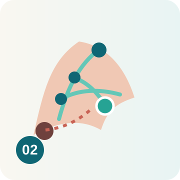
The true drainage pathway and nodal basins are defined. The Ukrainian protocol names technetium or indocyanine green for sentinel-node detection.
Ask: which mapping method was used?
The node or nodes should correspond to the lymphatic map and the criteria of the chosen navigation method.
Ask: how many sentinel nodes were found and in which basins?

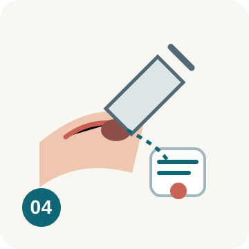
Each sentinel node should be clearly identifiable to pathology by location.
Ask: how many nodes/containers were submitted and how were they labelled?
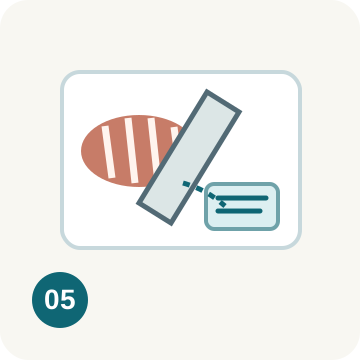
The Ukrainian protocol specifies nodal tissue pieces no more than 2-3 mm in paraffin blocks. The aim is adequate tissue coverage.
Ask: was the whole sentinel node processed using a dedicated protocol?
Ukrainian minimum: at least 3 levels, H&E and IHC, and at least 3 melanocyte-specific antibodies. Exact level spacing follows the validated laboratory protocol.
Ask: which levels and IHC markers were used?
Number examined and positive, largest deposit size, location, extranodal extension and other relevant tumour-burden descriptors.
Ask: is the report sufficient for accurate pN staging and management?
What happens to the sentinel node after removal
Removal of the sentinel lymph node is only part of the procedure. An equally important stage follows: appropriate gross processing, serial sectioning, morphologic assessment and immunohistochemistry according to protocol.

A checklist is not a legal conclusion
Missing detail in a short report does not prove that a step was omitted. First obtain the full operative note, mapping/nuclear-medicine documentation and pathology records.