What should a sentinel-node pathology report contain?
“No metastasis identified” or “sentinel node positive” gives the final answer, but may not show enough detail to assess tumour burden or the completeness of the pathology process.
What protocol No.1064 explicitly requires
These are the national minimum reporting fields after sentinel-node assessment.
- Sentinel-node location
- Regional basin / side.
- Number examined
- Total number of sentinel nodes submitted and assessed.
- Number positive
- Number containing confidently identified melanoma metastasis.
- Number and location of metastases
- Description of metastatic deposits within the node.
- Largest deposit
- Maximum dimension in millimetres.
- Extranodal extension
- Presence or absence.
- Conglomerates / other nodal descriptors
- Reported as applicable to the material and protocol.
What is useful in a high-quality report beyond the minimum
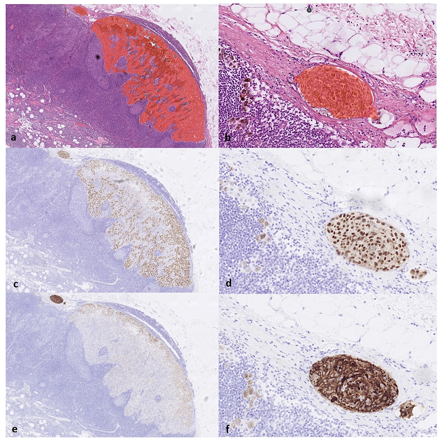
EORTC recommends more detailed characterization of microanatomic tumour burden.
Subcapsular, parenchymal, combined or other pattern.
Describes the structure of tumour burden rather than binary positivity alone.
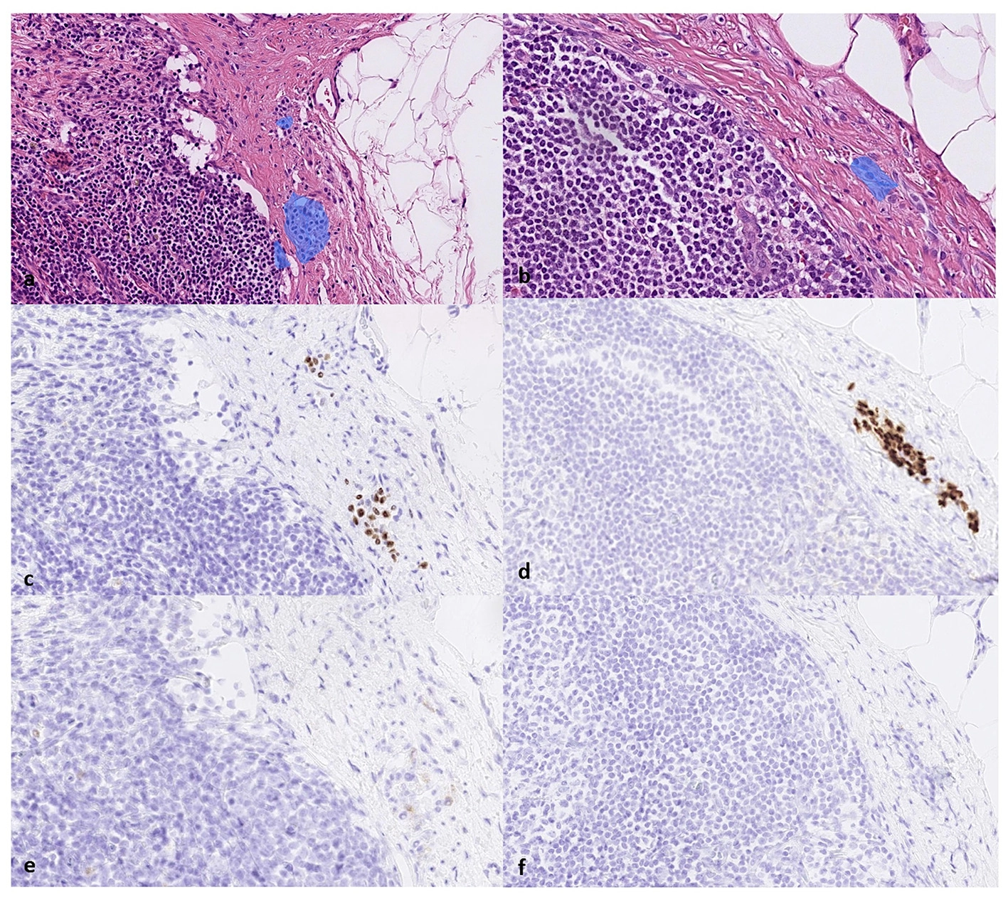
Useful to document when present, particularly in difficult differential diagnoses.
H&E and/or IHC in morphologic context; transparent documentation helps expert review.
A short report is not automatically wrong, but may be insufficient for quality audit
Missing detail in a discharge summary does not prove the laboratory failed to assess it. Distinguish a short clinical summary from the full pathology report.
“Sentinel lymph node: no melanoma metastasis identified.”
This communicates the final answer but does not show the number of sentinel nodes, the processing method, levels or IHC.
Left axillary sentinel nodes: 2 examined. Positive 1/2. Largest metastatic deposit 0.4 mm, subcapsular. No extranodal extension. H&E + IHC panel interpreted in morphologic context. Nodal nevus: absent / document if present.
What matters separately from the final wording
Not every technical parameter is necessarily a literal mandatory report field, but it is critical for expert quality review.
| Question | Why it matters |
|---|---|
| How was the node grossed? | The Ukrainian protocol specifies 2-3 mm tissue pieces in paraffin blocks. |
| How many levels were examined? | Ukrainian minimum ≥3; exact spacing follows a validated laboratory protocol. |
| Which IHC markers were used? | The Ukrainian protocol requires at least 3 melanocyte-specific antibodies. |
| Was morphology correlated? | Positive IHC alone does not prove metastasis, particularly when nodal nevus is a consideration. |
What to collect before a pathology second opinion
Review is most useful when the expert has more than a single-page report.
Breslow thickness, ulceration, mitotic activity and other key features.
Not just a short discharge-summary entry.
For repeat morphology and additional IHC when needed.
To correlate the number and location of removed nodes with the actual mapping procedure.