SLNB in melanoma: procedural and pathology quality standards
From indication and lymphatic mapping to grossing, serial levels, immunohistochemistry and pN staging. The central principle is that SLNB quality is determined by the entire multidisciplinary process, not a single step.
Plan before wide excision changes lymphatic drainage
SLNB pathologically stages clinically node-negative regional basins. Indications depend on the risk of occult nodal disease.
| Situation | Ukraine, protocol No.1064 | ESMO / ASCO-SSO | Practical point |
|---|---|---|---|
| pT1a <0.8 mm without ulceration | Not routinely recommended. | Not routine; may be discussed in selected high-risk situations. | Do not turn SLNB into an automatic procedure for thin melanoma. |
| pT1b | Recommended in the national protocol. | ESMO: discuss with the patient. | Shared decision-making with risk/benefit discussion. |
| Clinically N0, ≥1 mm | Used for pathological staging. | ESMO recommends for T2a and above. | Core SLNB setting in the absence of clinical nodal disease. |
| Wide local excision | SLNB should be performed together with WLE to avoid disturbing lymphatic drainage. | International practice likewise plans mapping before disruption of lymphatic pathways. | Timing is part of procedural quality. |
Sentinel-node identification methods in the current Ukrainian protocol
For sentinel-node detection, Ukrainian Unified Clinical Protocol No.1064 explicitly provides two approaches: technetium-based radionuclide mapping or indocyanine green (ICG). The aim is to reproduce the true lymphatic drainage pathway and identify all sentinel nodes.
| Method | Practical role | Key principle |
|---|---|---|
| Technetium-based radionuclide mapping | Preoperative lymphatic mapping and intraoperative node identification using a gamma probe. | Defines draining basins and sentinel nodes according to functional lymphatic drainage. |
| Indocyanine green (ICG) | Fluorescence-guided intraoperative navigation. | Also explicitly named in the current Ukrainian protocol as a method of sentinel-node detection. |
“One block - one section” is not an adequate sentinel-node protocol
The aim is to examine enough tissue volume that a small metastatic deposit is not left between two examined planes.

| Protocol | Gross/microscopic handling |
|---|---|
| Ukraine, No.1064 | Paraffin-block tissue pieces ≤2-3 mm; serial sections at at least 3 levels; H&E + IHC. |
| RCPA | Slice sentinel nodes at approximately 2-mm intervals along the long axis; submit all sections; step sections and IHC are used for assessment. |
| EORTC 2019 | Standardized multilevel sectioning; spacing is adapted to node size/shape. Minimum intervals may be around 50 μm, while other validated protocols use wider spacing. |
| CAP | Does not impose one universal numeric level algorithm for every sentinel node; adequate tissue assessment and complete reporting remain central. |
IHC increases sensitivity but does not replace morphology
The Ukrainian protocol requires at least three melanocyte-specific antibodies. Examples include SOX10, S100, HMB45, Melan-A, tyrosinase and MITF.
| Marker / situation | Practical interpretation |
|---|---|
| SOX10 / S100 | Sensitive melanocytic markers, but may also be positive in nodal nevi. |
| Melan-A | Supports melanocytic differentiation; benign nevus cells may also stain. |
| HMB45 | May help with distinction, but sensitivity varies. |
| PRAME / p16 | Useful adjuncts in difficult cases; neither is sufficient alone. Negative PRAME does not exclude melanoma. |
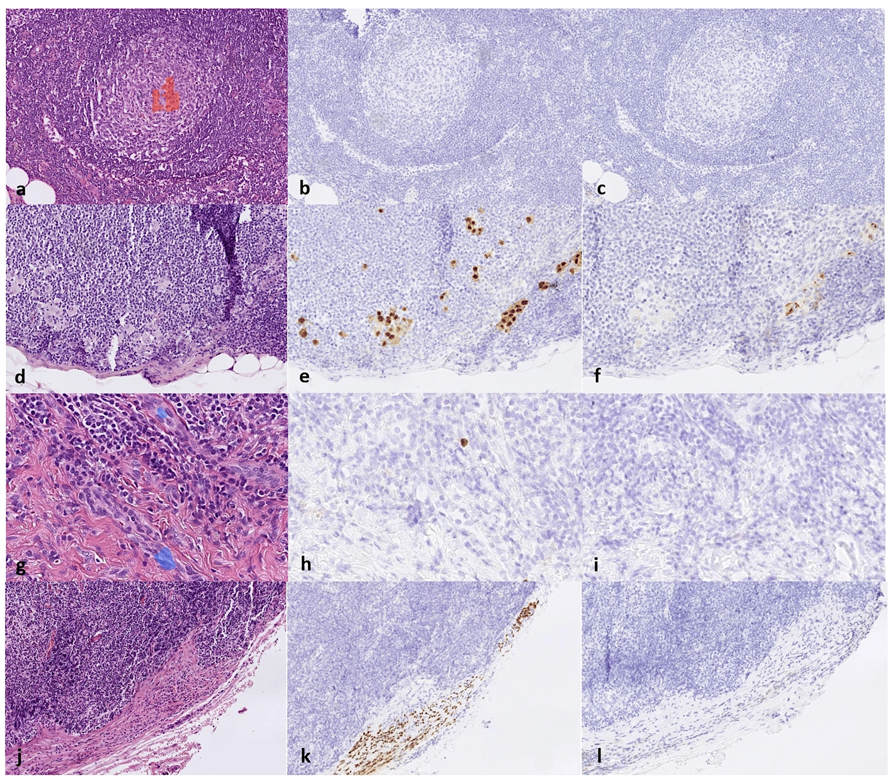
| Isolated IHC-positive cell | Without morphologic features of melanoma, should not automatically render the sentinel node positive. |
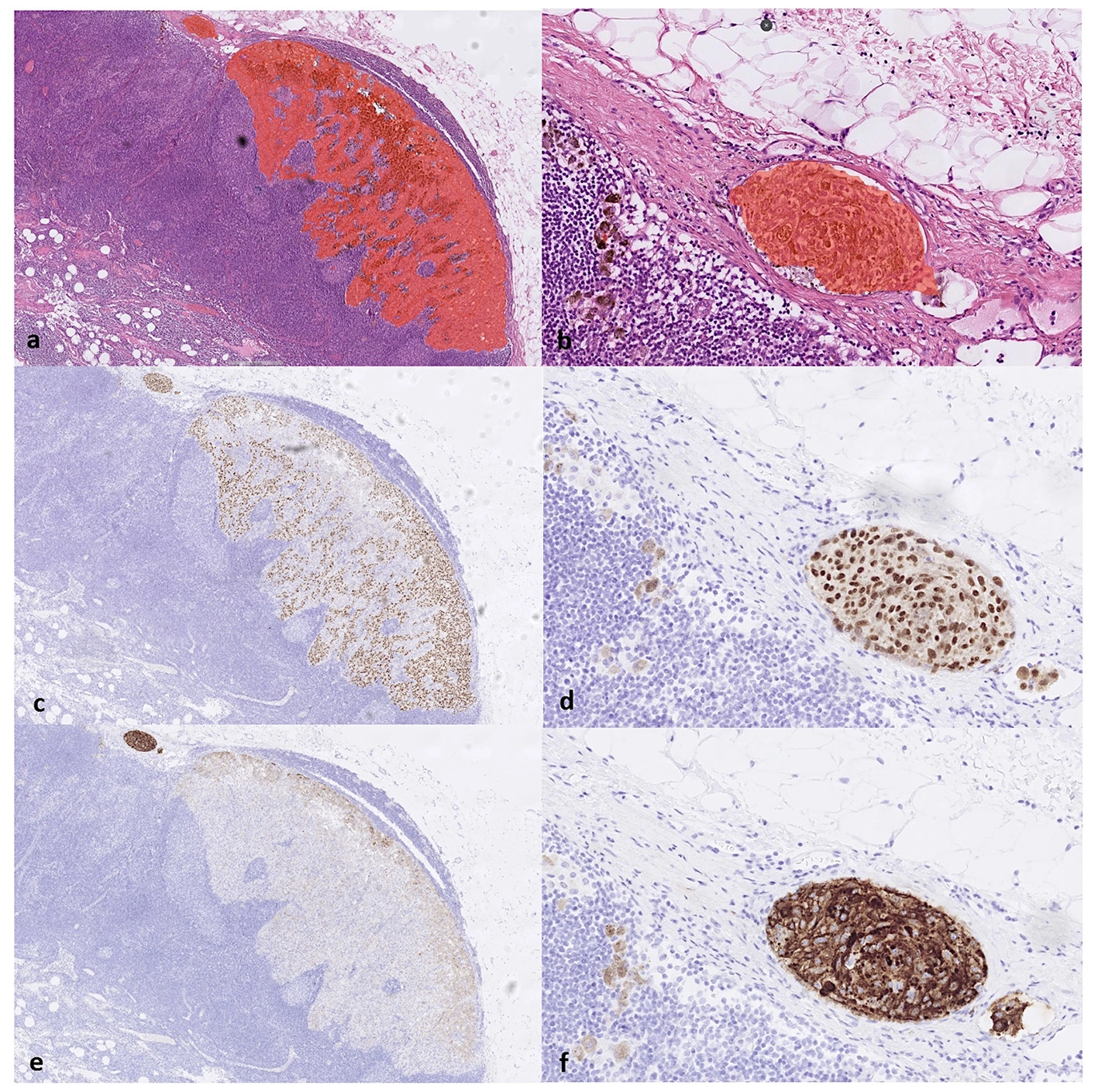
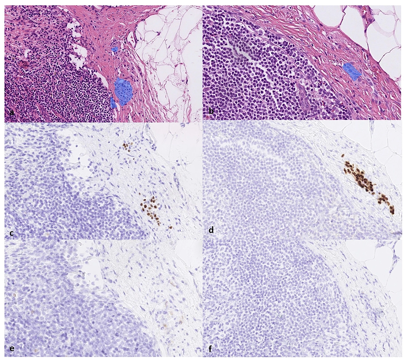
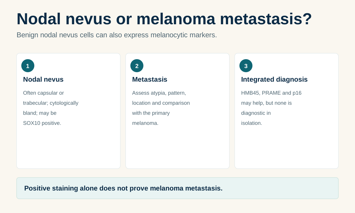
More than “metastasis present / absent”
The Ukrainian protocol already requires multiple quantitative and topographic fields; EORTC adds further detail on microanatomic tumour burden.
- Sentinel-node location
- Regional basin / side.
- Number of sentinel nodes
- How many nodes were actually examined.
- Number positive
- How many contain confidently identified melanoma metastasis.
- Number and location of deposits
- Subcapsular, parenchymal, combined or other pattern.
- Largest deposit
- Maximum dimension in millimetres.
- Extranodal extension
- Present/absent.
- Nodal nevus cells
- EORTC recommends noting them, particularly in difficult differential diagnoses.
- Method of detection
- H&E and/or IHC interpreted in morphologic context.
A false-negative result can arise at different stages
False-negative SLNB should not be reduced to pathology alone. Failure may occur during mapping, surgery or laboratory evaluation.
An unrecognized basin, interval node or incomplete lymphatic map.
Failure to remove one of the sentinel nodes or misinterpretation of navigation.
Insufficient sectioning, a missed small deposit or incorrect IHC interpretation.
Examples of diagnostic pitfalls
External figures are used only when reuse rights are explicit. The key pathology figures here are CC BY.
Evidence backbone
The page separates the Ukrainian regulatory framework, guideline recommendations and observational/meta-analytic evidence.